Terry Fox Laboratory, British Columbia Cancer, Vancouver, BC, Canada.
Interdisciplinary Oncology Program, University of British Columbia (UBC), Vancouver, BC, Canada.
Front Immunol. 2023 Sep 22;14:1198310. doi: 10.3389/fimmu.2023.1198310. eCollection 2023.
The three groups of helper innate lymphoid cells (ILCs), namely ILC1, ILC2 and ILC3, have been identified by flow cytometry by combinations of cell surface markers. Here, we review various ways ILCs are currently identified, focusing on potential problems and their solutions. The first step to identify all ILCs is to exclude other lymphocytes and myeloid cells by their lineage-specific markers (Lin). However, the Lin cocktail varies in various studies, and the definition of Lin- population containing ILCs is often ambiguous, resulting in contamination of Lin cells, particularly T cells.
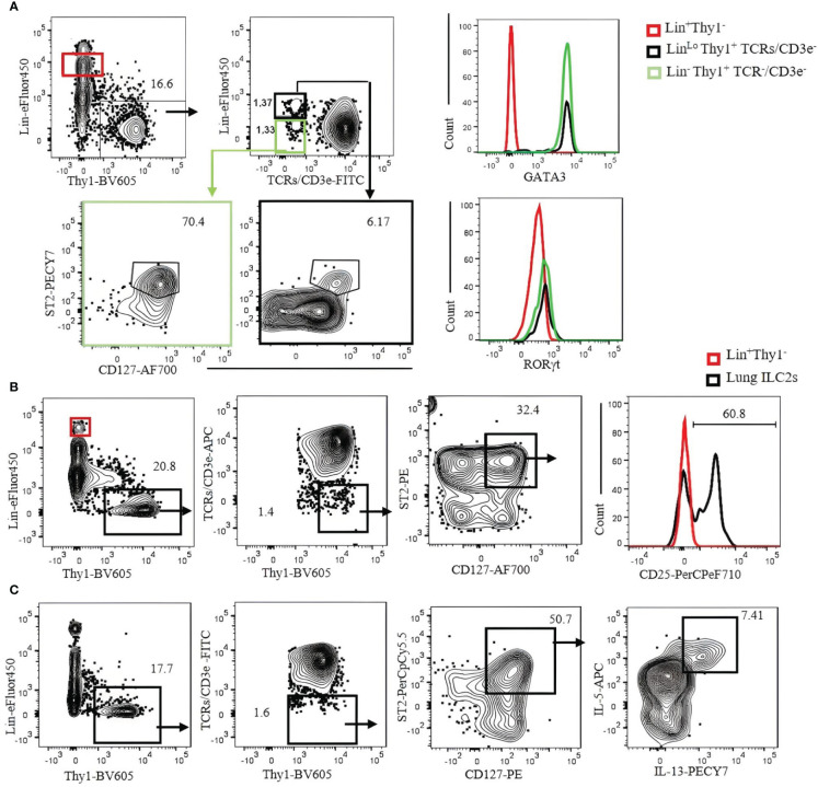
We have designed combinations of cell surface markers to identify ILC populations in various tissues of B6 mice by flow cytometry. To minimize T cell contamination, TCR/CD3ϵ antibodies were used separately from the Lin cocktail. ILCs identified by surface markers are confirmed by the expression of the transcription factors GATA3, RORγt, T-bet and Eomes.
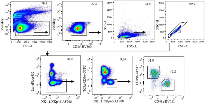
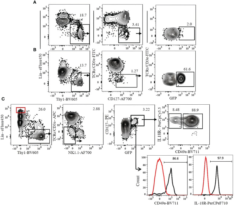
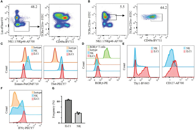
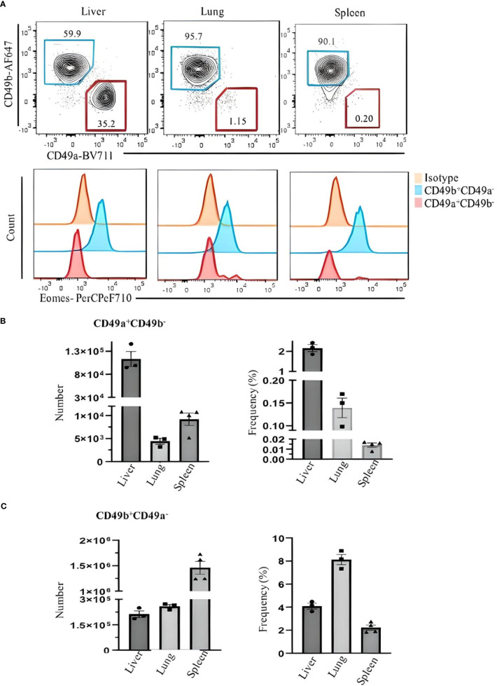
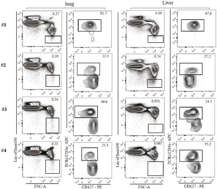
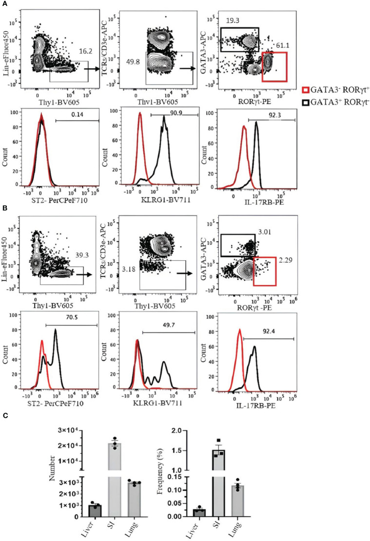
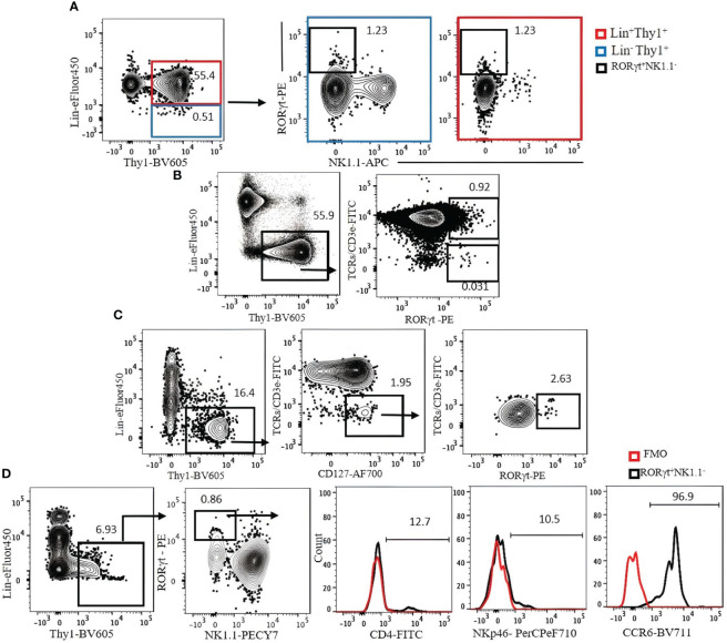
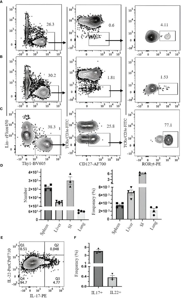
ILC1s in the B6 mouse liver are identified by Lin-NKp46NK1.1TCR/CD3ϵCD49aCD49b. However, defining ILC1s in other tissues remains a challenge. ILC2s in the lung are identified by LinTCR/CD3ϵ Thy1CD127ST2 whereas ILC2s in the small intestine and liver are identified by LinTCR/CD3ϵThy1GATA3RORγt. ILC3s in B6 mouse spleen, liver, lung and small intestine are identified by LinTCR/CD3ϵ Thy1CD127RORγt.
The ILC population is heterogeneous and the strategies to identify ILCs have to be designed for each ILC population and tissue. Excluding T cells in all cases is crucial, and a combination of transcription factors GATA3, RORγt, T-bet, and Eomes should be used to identify ILCs. Using CD3ϵ/TCRs in a different fluorochrome not in Lin cocktail minimizes contamination of T cells specifically identify individual ILC populations in various tissues.
通过细胞表面标志物的组合,已经鉴定出了三种辅助先天淋巴样细胞(ILC)组,即 ILC1、ILC2 和 ILC3。在这里,我们回顾了目前鉴定 ILC 的各种方法,重点介绍了潜在的问题及其解决方案。鉴定所有 ILC 的第一步是通过其谱系特异性标志物(Lin)排除其他淋巴细胞和髓样细胞。然而,Lin 鸡尾酒在不同的研究中有所不同,并且包含 ILC 的 Lin-群体的定义常常不明确,导致 Lin 细胞,特别是 T 细胞的污染。
我们通过流式细胞术设计了组合细胞表面标志物来鉴定 B6 小鼠各种组织中的 ILC 群体。为了最大程度地减少 T 细胞污染,TCR/CD3ε 抗体与 Lin 鸡尾酒分开使用。通过转录因子 GATA3、RORγt、T-bet 和 Eomes 的表达来确认通过表面标志物鉴定的 ILC。
B6 小鼠肝脏中的 ILC1 由 Lin-NKp46NK1.1TCR/CD3εCD49aCD49b 鉴定。然而,鉴定其他组织中的 ILC1 仍然是一个挑战。肺中的 ILC2 由 Lin-TCR/CD3εThy1CD127ST2 鉴定,而小肠和肝脏中的 ILC2 由 Lin-TCR/CD3εThy1GATA3RORγt 鉴定。B6 小鼠脾、肝、肺和小肠中的 ILC3 由 Lin-TCR/CD3εThy1CD127RORγt 鉴定。
ILC 群体是异质的,鉴定 ILC 的策略必须针对每个 ILC 群体和组织进行设计。在所有情况下排除 T 细胞至关重要,并且应该使用转录因子 GATA3、RORγt、T-bet 和 Eomes 的组合来鉴定 ILC。在 Lin 鸡尾酒中不使用不同荧光染料的 CD3ε/TCRs 可以最大程度地减少 T 细胞的污染,专门鉴定各种组织中的单个 ILC 群体。