Laboratory of Biomedical Optics and Applied Biophysics, School of Electrical and Computer Engineering, National Technical University of Athens, Zografou Campus, 15780, Athens, Greece.
Division of Pharmaceutical Technology, School of Pharmacy, National and Kapodistrian University of Athens, Panepistimioupoli, Zografou Campus, 15771, Athens, Greece.
Ann Biomed Eng. 2024 Feb;52(2):376-385. doi: 10.1007/s10439-023-03384-x. Epub 2023 Oct 18.
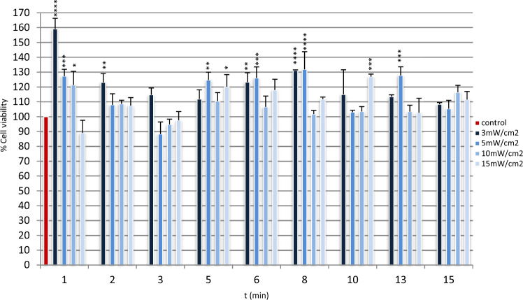
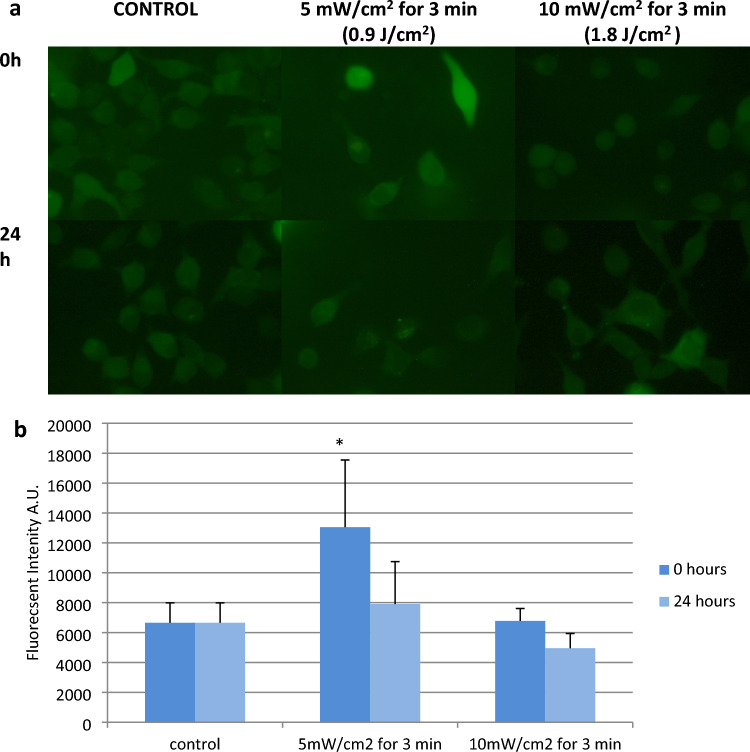
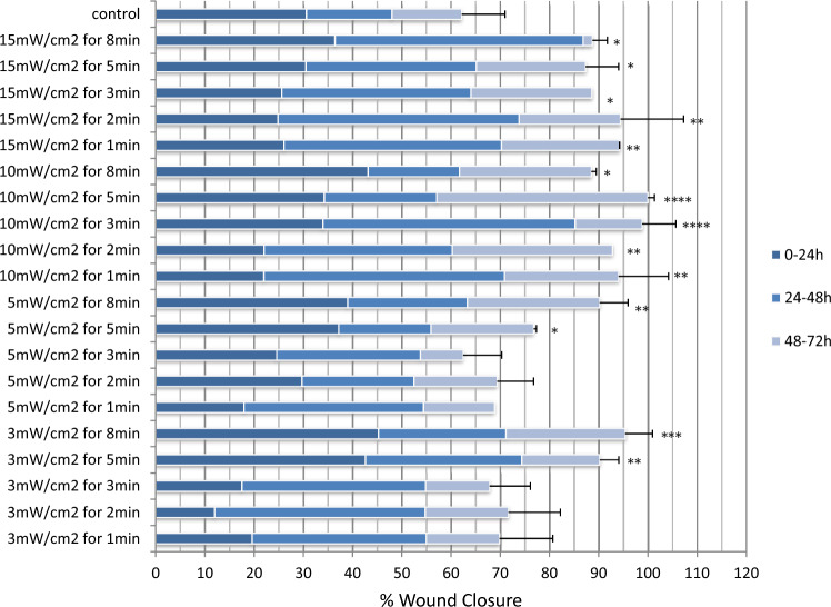
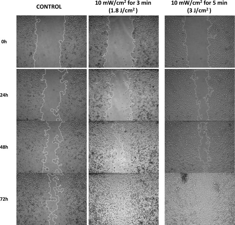
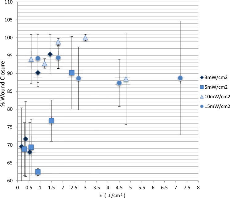
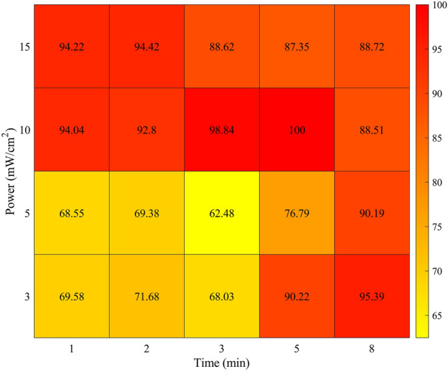
This study aims to investigate the effectiveness of low power red light (661 nm) in accelerating the wound healing process of an in vitro scratch assay model of keratinocytes. Furthermore, the study aims to clarify the role of light irradiation parameters, optimize them and gain additional insight into the mechanisms of wound closure as a result of photobiomodulation. Wound healing was studied using scratch assay model of NCTC 2544 keratinocytes. Cells were irradiated with a laser at various power densities and times. Images were acquired at 0, 24, 48 and 72 h following the laser treatment. Cellular proliferation was studied by MTT. ROS were studied at 0 and 24 h by fluorescence microscopy. Image analysis was used to determine the wound closure rates and quantify ROS. The energy range of 0.18-7.2 J/cm was not phototoxic, increased cell viability and promoted wound healing. Power and irradiation time proved to be more important than energy. The results indicated the existence of two thresholds in both power and irradiation time that need to be overcome to improve wound healing. An increase in ROS production was observed at 0 h only in the group with the lowest healing rate. This early response seemed to block proliferation and finally wound healing. Low level laser light at 661 nm enhanced both proliferation and migration in keratinocytes, providing evidence that it could possibly stimulate wound healing in vivo. The observed results are dependent on irradiance and irradiation time rather than energy dose in total.
本研究旨在探讨低功率红光(661nm)对体外角质形成细胞划痕试验模型的伤口愈合过程的加速作用。此外,本研究旨在阐明光辐照参数的作用,优化这些参数,并深入了解光生物调节导致伤口闭合的机制。 使用 NCTC 2544 角质形成细胞的划痕试验模型研究伤口愈合。细胞在不同的功率密度和时间下用激光照射。在激光处理后 0、24、48 和 72 小时采集图像。通过 MTT 研究细胞增殖。在 0 和 24 小时通过荧光显微镜研究 ROS。使用图像分析来确定伤口闭合率并量化 ROS。能量范围为 0.18-7.2J/cm 时不会产生光毒性,反而会增加细胞活力并促进伤口愈合。功率和辐照时间被证明比能量更重要。结果表明,在提高伤口愈合率方面,功率和辐照时间都存在两个需要克服的阈值。仅在愈合率最低的组中观察到在 0 小时时 ROS 产量增加。这种早期反应似乎会阻止增殖,最终阻止伤口愈合。661nm 的低水平激光光增强了角质形成细胞的增殖和迁移,这表明它可能在体内刺激伤口愈合。观察到的结果取决于辐照度和辐照时间,而不是总能量剂量。