Shalaby Manal, Hamouda Dalia, Khedr Shaimaa M, Mostafa Haitham M, Saeed Hesham, Ghareeb Ahmed Z
Medical Biotechnology Department, Institute of Genetic Engineering and Biotechnology, City of Scientific Research and Technological Applications, Alexandria, Egypt.
Centre of Excellence for Drug Preclinical Studies (CE-DPS), Pharmaceutical and Fermentation Industry Development Centre, City of Scientific Research and Technological Applications, New Borg El Arab, Alexandria, Egypt.
PLoS One. 2023 Oct 20;18(10):e0282557. doi: 10.1371/journal.pone.0282557. eCollection 2023.
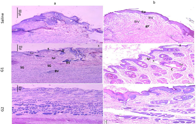
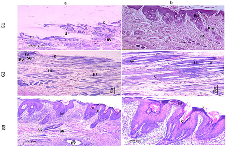
The creation of innovative wound-healing nanomaterials based on natural compounds emerges as a top research goal. This research aimed to create a gel containing collagen nanoparticles and evaluate its therapeutic potential for skin lesions. Collagen nanoparticles were produced from fish scales using desolvation techniques. Using SDS PAGE electrophoresis, Fourier transform infrared spectroscopy (FTIR) as well as the structure of the isolated collagen and its similarities to collagen type 1 were identified. The surface morphology of the isolated collagen and its reformulation into nanoparticles were examined using transmission and scanning electron microscopy. A Zeta sizer was used to examine the size, zeta potential, and distribution of the synthesized collagen nanoparticles. The cytotoxicity of the nanomaterials was investigated and an experimental model was used to evaluate the wound healing capability. The overall collagen output from Tilapia fish scales was 42%. Electrophoretic patterns revealed that the isolated collagen included a unique protein with chain bands of 126-132 kDa and an elevated beta band of 255 kDa. When compared to the isolated collagen, the collagen nanoparticles' FTIR results revealed a significant drop in the amide II (42% decrease) and amide III (32% decrease) band intensities. According to SEM analysis, the generated collagen nanoparticles ranged in size from 100 to 350 nm, with an average diameter of 182 nm determined by the zeta sizer. The produced collagen nanoparticles were polydispersed in nature and had an equivalent average zeta potential of -17.7 mV. Cytotoxicity study showed that, when treating fibroblast cells with collagen nanoparticle concentrations, very mild morphological alterations were detected after human skin fibroblasts were treated with collagen nanoparticles 32 μg/ml for 24 hours, as higher concentrations of collagen nanoparticles caused cell detachment. Macroscopical and histological investigations proved that the fabricated fish scale collagen nanoparticles promoted the healing process in comparison to the saline group.
基于天然化合物的创新型伤口愈合纳米材料的研发成为首要研究目标。本研究旨在制备一种含有胶原蛋白纳米颗粒的凝胶,并评估其对皮肤损伤的治疗潜力。采用去溶剂化技术从鱼鳞中制备胶原蛋白纳米颗粒。通过SDS-PAGE电泳、傅里叶变换红外光谱(FTIR)鉴定了分离出的胶原蛋白的结构及其与I型胶原蛋白的相似性。利用透射电子显微镜和扫描电子显微镜检查了分离出的胶原蛋白的表面形态及其重新形成纳米颗粒的情况。使用Zeta粒度分析仪检测合成的胶原蛋白纳米颗粒的尺寸、Zeta电位和分布。研究了纳米材料的细胞毒性,并使用实验模型评估伤口愈合能力。罗非鱼鱼鳞的总胶原蛋白产量为42%。电泳图谱显示,分离出的胶原蛋白包含一种独特的蛋白质,其链带为126-132 kDa,β带升高至255 kDa。与分离出的胶原蛋白相比,胶原蛋白纳米颗粒的FTIR结果显示酰胺II(降低42%)和酰胺III(降低32%)带强度显著下降。根据扫描电子显微镜分析,生成的胶原蛋白纳米颗粒尺寸在100至350 nm之间,Zeta粒度分析仪测定的平均直径为182 nm。所制备的胶原蛋白纳米颗粒在本质上是多分散的,平均Zeta电位为-17.7 mV。细胞毒性研究表明,在用胶原蛋白纳米颗粒浓度处理成纤维细胞时,当人皮肤成纤维细胞用32 μg/ml胶原蛋白纳米颗粒处理24小时后,检测到非常轻微的形态改变,因为更高浓度的胶原蛋白纳米颗粒会导致细胞 detachment。宏观和组织学研究证明,与生理盐水组相比,所制备的鱼鳞胶原蛋白纳米颗粒促进了愈合过程。