Department of Cardiology, West China Hospital, Sichuan University, Guoxue Xiang No. 37, Guo Xue Road, Chengdu, 610041, Sichuan, People's Republic of China.
Department of Radiology, West China Hospital, Sichuan University, Guoxue Xiang No. 37, Chengdu, 610041, Sichuan, People's Republic of China.
J Cardiovasc Magn Reson. 2023 Nov 16;25(1):64. doi: 10.1186/s12968-023-00974-5.
Although reference ranges of T1 and T2 mapping are well established for cardiovascular magnetic resonance (CMR) at 1.5T, data for 3T are still lacking. The objective of this study is to establish reference ranges of myocardial T1 and T2 based on a large multicenter cohort of healthy Chinese adults at 3T CMR.
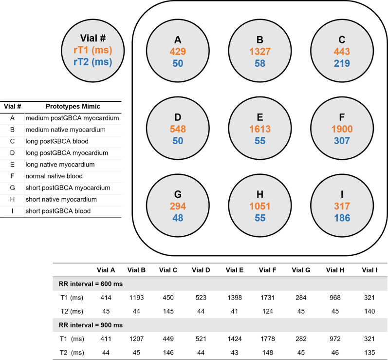
A total of 1015 healthy Chinese adults (515 men, age range: 19-87 years) from 11 medical centers who underwent CMR using 3T Siemens scanners were prospectively enrolled. T1 mapping was performed with a motion-corrected modified Look-Locker inversion recovery sequence using a 5(3)3 scheme. T2 mapping images were acquired using T2-prepared fast low-angle shot sequence. T1 and T2 relaxation times were quantified for each slice and each myocardial segment. The T1 mapping and extracellular volume standardization (T1MES) phantom was used for quality assurance at each center prior to subject scanning.
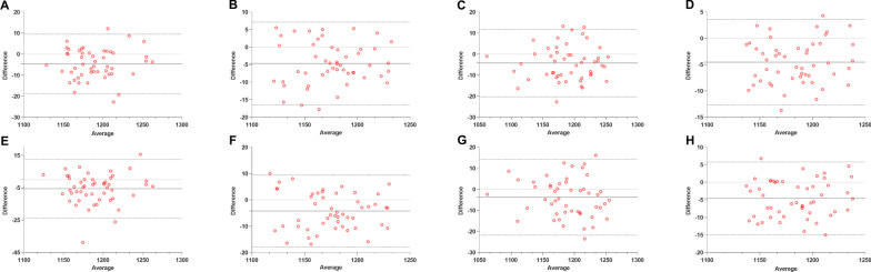
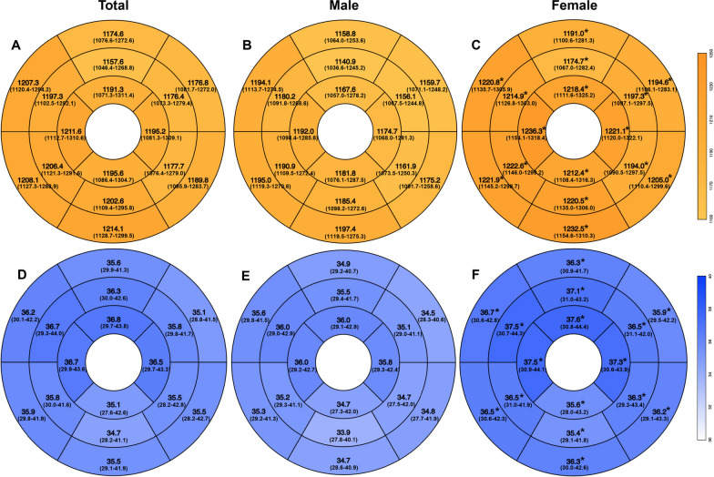
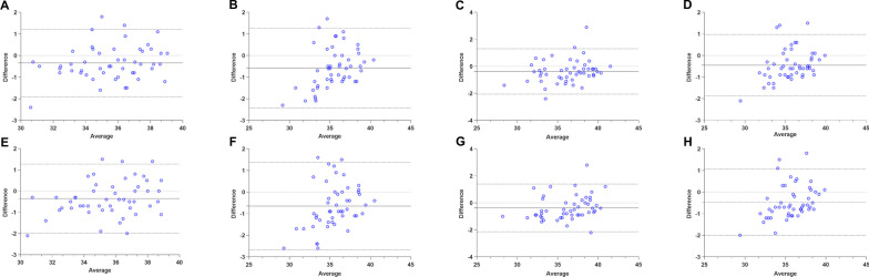
The phantom analysis showed strong consistency of spin echo, T1 mapping, and T2 mapping among centers. In the entire cohort, global T1 and T2 reference values were 1193 ± 34 ms and 36 ± 2.5 ms. Global T1 and T2 values were higher in females than in males (T1: 1211 ± 29 ms vs. 1176 ± 30 ms, p < 0.001; T2: 37 ± 2.3 ms vs. 35 ± 2.5 ms, p < 0.001). There were statistical differences in global T2 across age groups (p < 0.001), but not in global T1. Linear regression showed no correlation between age and global T1 or T2 values. In males, positive correlation was found between heart rate and global T1 (r = 0.479, p < 0.001).
Using phantom-validated imaging sequences, we provide reference ranges for myocardial T1 and T2 values on 3T scanners in healthy Chinese adults, which can be applied across participating sites. Trial registration URL: http://www.chictr.org.cn/index.aspx . Unique identifier: ChiCTR1900025518. Registration name: 3T magnetic resonance myocardial quantitative imaging standardization and reference value study: a multi-center clinical study.
尽管心血管磁共振(CMR)在 1.5T 场强下已经建立了 T1 和 T2 弛豫时间的参考范围,但在 3T 场强下的数据仍然缺乏。本研究的目的是基于在 3T CMR 检查的大型多中心中国健康成年人队列,建立心肌 T1 和 T2 的参考范围。
前瞻性纳入了来自 11 家医疗中心的 1015 名健康中国成年人(515 名男性,年龄范围:19-87 岁),他们使用 3T 西门子扫描仪进行了 CMR 检查。T1 映射使用带运动校正的改良 Look-Locker 反转恢复序列,采用 5(3)3 方案进行。T2 映射图像使用 T2 准备的快速低角度 shot 序列采集。对每个切片和每个心肌节段定量分析 T1 和 T2 弛豫时间。在对受试者进行扫描之前,每个中心都使用 T1 映射和细胞外容积标准化(T1MES)体模进行质量保证。
体模分析显示各中心间自旋回波、T1 映射和 T2 映射具有很强的一致性。在整个队列中,全球 T1 和 T2 的参考值分别为 1193±34ms 和 36±2.5ms。女性的全球 T1 和 T2 值高于男性(T1:1211±29ms vs. 1176±30ms,p<0.001;T2:37±2.3ms vs. 35±2.5ms,p<0.001)。各年龄组的全球 T2 存在统计学差异(p<0.001),但全球 T1 无差异。线性回归显示年龄与全球 T1 或 T2 值无相关性。在男性中,心率与全球 T1 呈正相关(r=0.479,p<0.001)。
使用经过体模验证的成像序列,我们提供了健康中国成年人在 3T 扫描仪上心肌 T1 和 T2 值的参考范围,可在参与研究的各个中心应用。试验注册网址:http://www.chictr.org.cn/index.aspx. 唯一标识符:ChiCTR1900025518. 注册名称:3T 磁共振心肌定量成像标准化及参考值研究:多中心临床研究。