• 文献检索
  • 文档翻译
  • 深度研究
  • 学术资讯
  • Suppr Zotero 插件Zotero 插件
  • 邀请有礼
  • 套餐&价格
  • 历史记录
应用&插件
Suppr Zotero 插件Zotero 插件浏览器插件Mac 客户端Windows 客户端微信小程序
定价
高级版会员购买积分包购买API积分包
服务
文献检索文档翻译深度研究API 文档MCP 服务
关于我们
关于 Suppr公司介绍联系我们用户协议隐私条款
关注我们

Suppr 超能文献

核心技术专利:CN118964589B侵权必究
粤ICP备2023148730 号-1Suppr @ 2026

文献检索

告别复杂PubMed语法,用中文像聊天一样搜索,搜遍4000万医学文献。AI智能推荐,让科研检索更轻松。

立即免费搜索

文件翻译

保留排版,准确专业,支持PDF/Word/PPT等文件格式,支持 12+语言互译。

免费翻译文档

深度研究

AI帮你快速写综述,25分钟生成高质量综述,智能提取关键信息,辅助科研写作。

立即免费体验

基于人工智能的 3D MRI 分析在早期膝关节骨关节炎中,AI 确定的 Kellgren-Lawrence 分级与内侧半月板突出和软骨厚度的相关性。

Association of AI-determined Kellgren-Lawrence grade with medial meniscus extrusion and cartilage thickness by AI-based 3D MRI analysis in early knee osteoarthritis.

机构信息

Center for Stem Cell and Regenerative Medicine, Tokyo Medical and Dental University, Tokyo, Japan.

Center for Stem Cell and Regenerative Medicine, Tokyo Medical and Dental University (TMDU), 1-5-45 Yushima, Bunkyo-ku, Tokyo, 113-8510, Japan.

出版信息

Sci Rep. 2023 Nov 16;13(1):20093. doi: 10.1038/s41598-023-46953-9.

DOI:10.1038/s41598-023-46953-9
PMID:37973855
原文链接:https://pmc.ncbi.nlm.nih.gov/articles/PMC10654518/
Abstract

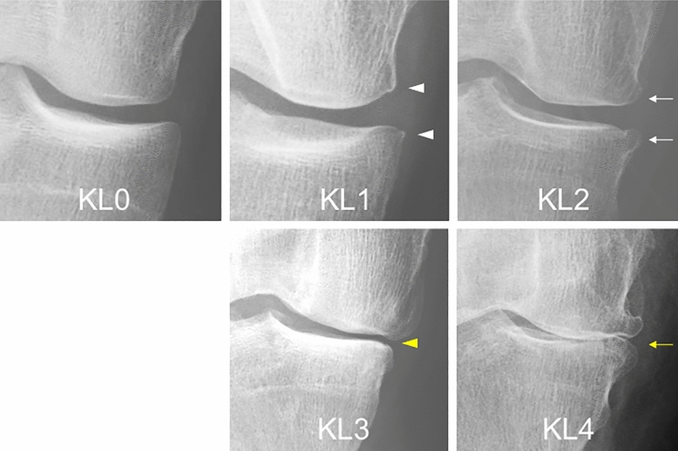
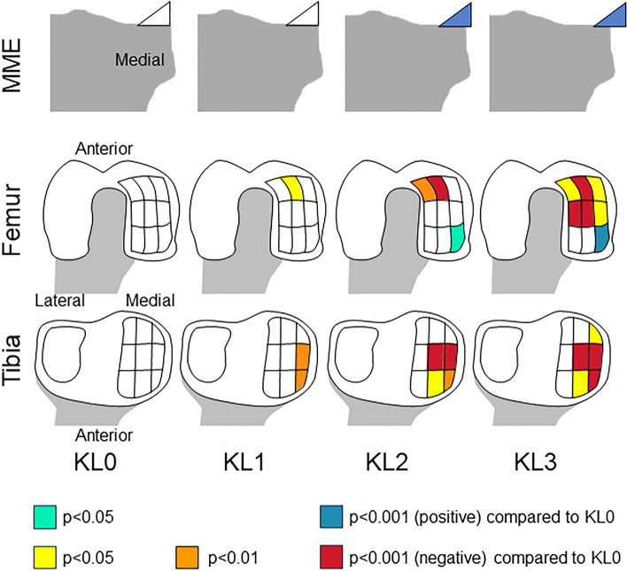
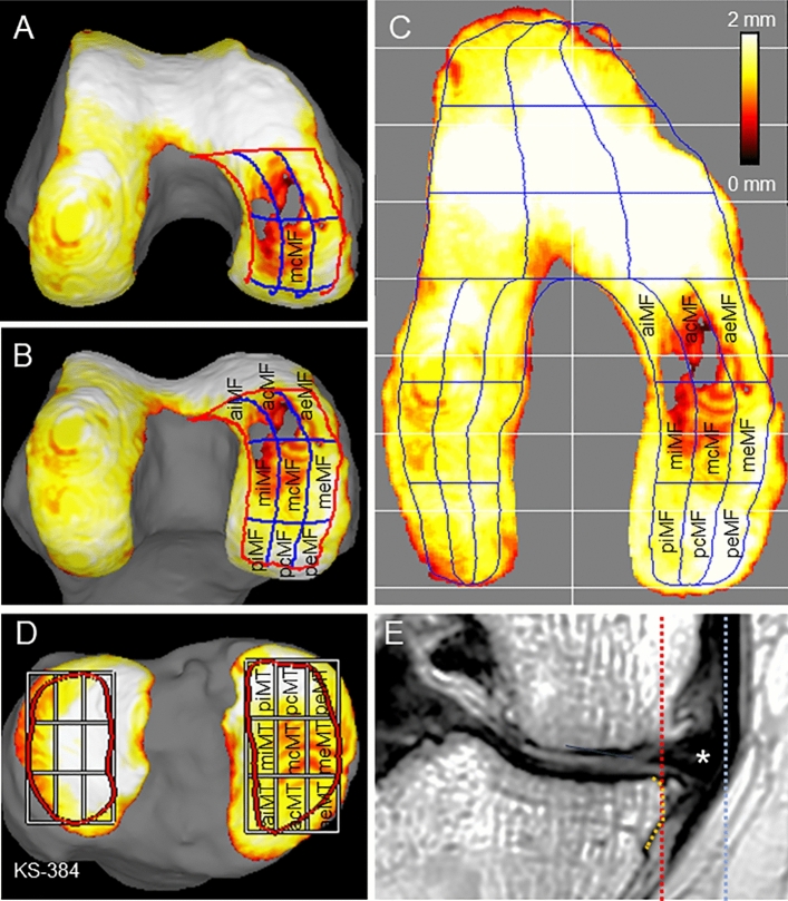
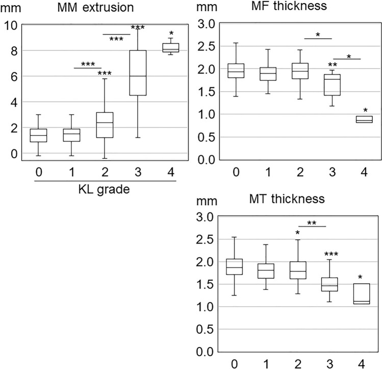
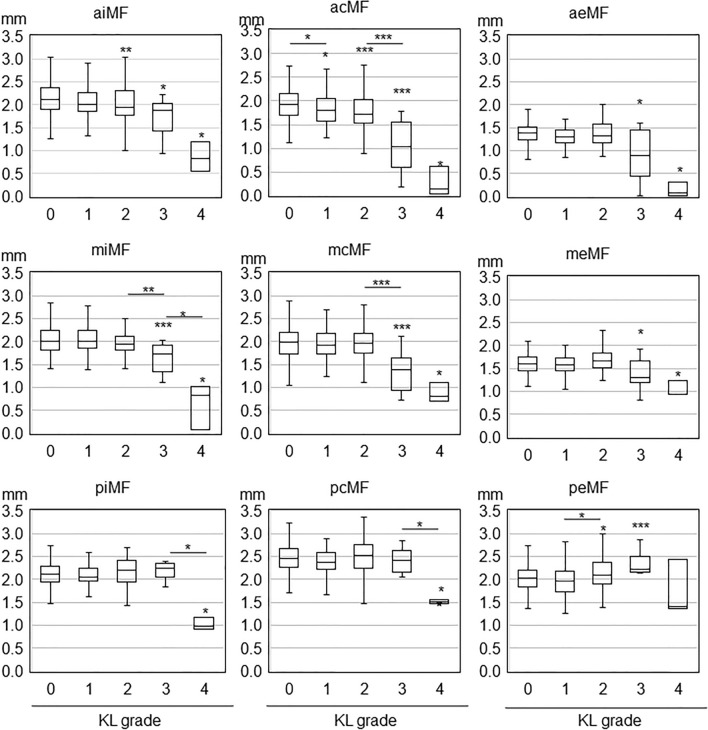
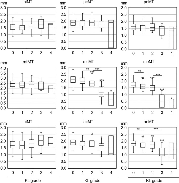
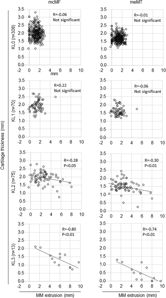
The associations among Kellgren-Lawrence (KL) grade, medial meniscus extrusion (MME), and cartilage thickness in knee osteoarthritis (OA) remain insufficiently understood. Our aim was to determine these associations in early to moderate medial tibiofemoral knee OA. We included 469 subjects with no lateral OA from the Kanagawa Knee Study. KL grade was assessed using artificial intelligence (AI) software. The MME was measured by MRI, and the cartilage thickness was evaluated in 18 subregions of the medial femorotibial joint by another AI system. The median MME width was 1.4 mm in KL0, 1.5 mm in KL1, 2.4 mm in KL2, and 6.0 mm in KL3. Cartilage thinning in the medial femur occurred in the anterior central subregion in KL1, expanded inwardly in KL2, and further expanded in KL3. Cartilage thinning in the medial tibia occurred in the anterior and middle external subregions in KL1, expanded into the anterior and middle central subregions in KL2, and further expanded in KL3. The absolute correlation coefficient between MME width and cartilage thickness increased as the KL grade increased in some subregions. This study provides novel insights into the early stages of knee OA and potentially has implications for the development of early intervention strategies.

摘要

在膝骨关节炎(OA)中,Kellgren-Lawrence(KL)分级、内侧半月板突出(MME)和软骨厚度之间的关联仍未得到充分理解。我们的目的是确定早期至中度内侧胫股膝关节 OA 中的这些关联。我们纳入了来自神奈川膝关节研究的 469 名无外侧 OA 的受试者。KL 分级采用人工智能(AI)软件进行评估。MME 通过 MRI 测量,软骨厚度通过另一个 AI 系统评估内侧股骨胫骨关节的 18 个亚区。KL0 的中位数 MME 宽度为 1.4mm,KL1 为 1.5mm,KL2 为 2.4mm,KL3 为 6.0mm。KL1 中股骨内侧的前中央亚区出现软骨变薄,KL2 中向内扩展,KL3 中进一步扩展。KL1 中胫骨内侧的前外侧和中外部亚区出现软骨变薄,KL2 中向前后中央亚区扩展,KL3 中进一步扩展。在某些亚区中,MME 宽度和软骨厚度之间的绝对相关系数随着 KL 分级的增加而增加。本研究为膝关节 OA 的早期阶段提供了新的见解,并可能对早期干预策略的发展具有重要意义。

https://cdn.ncbi.nlm.nih.gov/pmc/blobs/b247/10654518/6d139916cef6/41598_2023_46953_Fig7_HTML.jpg
https://cdn.ncbi.nlm.nih.gov/pmc/blobs/b247/10654518/951d75bd73f9/41598_2023_46953_Fig1_HTML.jpg
https://cdn.ncbi.nlm.nih.gov/pmc/blobs/b247/10654518/13bce3bfdc8d/41598_2023_46953_Fig2_HTML.jpg
https://cdn.ncbi.nlm.nih.gov/pmc/blobs/b247/10654518/802845cd62d9/41598_2023_46953_Fig3_HTML.jpg
https://cdn.ncbi.nlm.nih.gov/pmc/blobs/b247/10654518/8cce2be94012/41598_2023_46953_Fig4_HTML.jpg
https://cdn.ncbi.nlm.nih.gov/pmc/blobs/b247/10654518/6e09168bd329/41598_2023_46953_Fig5_HTML.jpg
https://cdn.ncbi.nlm.nih.gov/pmc/blobs/b247/10654518/8aa60b4b5895/41598_2023_46953_Fig6_HTML.jpg
https://cdn.ncbi.nlm.nih.gov/pmc/blobs/b247/10654518/6d139916cef6/41598_2023_46953_Fig7_HTML.jpg

相似文献

1
Association of AI-determined Kellgren-Lawrence grade with medial meniscus extrusion and cartilage thickness by AI-based 3D MRI analysis in early knee osteoarthritis.基于人工智能的 3D MRI 分析在早期膝关节骨关节炎中,AI 确定的 Kellgren-Lawrence 分级与内侧半月板突出和软骨厚度的相关性。
Sci Rep. 2023 Nov 16;13(1):20093. doi: 10.1038/s41598-023-46953-9.
2
Medial Tibial Osteophyte Width Strongly Reflects Medial Meniscus Extrusion Distance and Medial Joint Space Width Moderately Reflects Cartilage Thickness in Knee Radiographs.膝关节 X 线片中,胫骨内侧骨赘宽度强烈反映内侧半月板挤出距离,而内侧关节间隙宽度适度反映软骨厚度。
J Magn Reson Imaging. 2022 Sep;56(3):824-834. doi: 10.1002/jmri.28079. Epub 2022 Jan 27.
3
Heterogeneity of cartilage damage in Kellgren and Lawrence grade 2 and 3 knees: the MOST study.Kellgren 和 Lawrence 分级 2 和 3 膝关节软骨损伤的异质性:MOST 研究。
Osteoarthritis Cartilage. 2022 May;30(5):714-723. doi: 10.1016/j.joca.2022.02.614. Epub 2022 Feb 22.
4
Articular cartilage surface roughness as an imaging-based morphological indicator of osteoarthritis: A preliminary investigation of osteoarthritis initiative subjects.关节软骨表面粗糙度作为骨关节炎基于成像的形态学指标:骨关节炎倡议受试者的初步研究。
J Orthop Res. 2017 Dec;35(12):2755-2764. doi: 10.1002/jor.23588. Epub 2017 May 23.
5
T1rho MRI of menisci in patients with osteoarthritis at 3 Tesla: a preliminary study.3特斯拉下骨关节炎患者半月板的T1rho磁共振成像:一项初步研究。
J Magn Reson Imaging. 2014 Sep;40(3):588-95. doi: 10.1002/jmri.24437. Epub 2013 Nov 4.
6
Relationship between knee alignment and T1ρ values of articular cartilage and menisci in patients with knee osteoarthritis.膝关节骨关节炎患者膝关节对线与关节软骨和半月板 T1ρ 值的关系。
Eur J Radiol. 2013 Nov;82(11):1946-52. doi: 10.1016/j.ejrad.2013.05.010. Epub 2013 Jun 13.
7
Stage-specific meniscal features predict progression of osteoarthritis of the knee: a retrospective cohort study using data from the osteoarthritis initiative.特定分期的半月板特征可预测膝关节骨关节炎的进展:一项基于骨关节炎倡议数据的回顾性队列研究。
BMC Musculoskelet Disord. 2019 Jan 22;20(1):33. doi: 10.1186/s12891-019-2413-7.
8
Relationship between medial meniscus extrusion and cartilage measurements in the knee by fully automatic three-dimensional MRI analysis.全自动三维 MRI 分析膝关节内侧半月板突出与软骨测量值的关系。
BMC Musculoskelet Disord. 2020 Nov 12;21(1):742. doi: 10.1186/s12891-020-03768-3.
9
T1rho MRI of menisci and cartilage in patients with osteoarthritis at 3T.3T 下骨关节炎患者半月板和软骨的 T1rho MRI。
Eur J Radiol. 2012 Sep;81(9):2329-36. doi: 10.1016/j.ejrad.2011.07.017. Epub 2011 Sep 9.
10
Quantitative CT of the knee in the IMI-APPROACH osteoarthritis cohort: Association of bone mineral density with radiographic disease severity, meniscal coverage and meniscal extrusion.膝关节定量 CT 在 IMI-APPROACH 骨关节炎队列中的应用:骨密度与放射学疾病严重程度、半月板覆盖度和半月板外突的相关性。
Bone. 2023 Mar;168:116673. doi: 10.1016/j.bone.2023.116673. Epub 2023 Jan 7.

引用本文的文献

1
Distribution of interscan measurement error in AI-based 3D MRI analysis of knee cartilage thickness in osteoarthritis.骨关节炎患者膝关节软骨厚度的基于人工智能的三维磁共振成像分析中扫描间测量误差的分布
PLoS One. 2025 Aug 7;20(8):e0329610. doi: 10.1371/journal.pone.0329610. eCollection 2025.
2
Same-model and cross-model variability in knee cartilage thickness measurements using 3D MRI systems.使用3D MRI系统测量膝关节软骨厚度时的同模型和跨模型变异性。
PLoS One. 2025 Jun 13;20(6):e0324912. doi: 10.1371/journal.pone.0324912. eCollection 2025.
3
3D-MRI analysis of cartilage thickness changes after PRP injection in medial knee osteoarthritis: A preliminary report.

本文引用的文献

1
Morphological analysis of three-dimensional MR images of patellofemoral joints in asymptomatic subjects.对无症状受试者髌股关节的三维磁共振图像进行形态学分析。
Sci Rep. 2023 Oct 5;13(1):16750. doi: 10.1038/s41598-023-42404-7.
2
Association between knee cartilage thickness determined by magnetic resonance imaging three-dimensional analysis and the International Cartilage Repair Society (ICRS) arthroscopic grade.膝关节磁共振三维分析测定的软骨厚度与国际软骨修复学会(ICRS)关节镜分级的相关性。
Knee. 2023 Jun;42:90-98. doi: 10.1016/j.knee.2023.02.005. Epub 2023 Mar 21.
3
Artificial-Intelligence-Aided Radiographic Diagnostic of Knee Osteoarthritis Leads to a Higher Association of Clinical Findings with Diagnostic Ratings.
富血小板血浆注射治疗膝内侧骨关节炎后软骨厚度变化的3D-MRI分析:初步报告
PLoS One. 2025 Apr 30;20(4):e0321067. doi: 10.1371/journal.pone.0321067. eCollection 2025.
4
Evaluating potential for AI automation of quantitative and semi-quantitative MRI scoring in arthritis, especially at the knee: a systematic literature review.评估人工智能在关节炎定量和半定量MRI评分自动化方面的潜力,尤其是膝关节:一项系统文献综述。
Skeletal Radiol. 2025 Mar 26. doi: 10.1007/s00256-025-04922-5.
5
A systematic review and meta-analysis examining alterations in medial meniscus extrusion and clinical outcomes following high tibial osteotomy.一项关于胫骨高位截骨术后内侧半月板挤压变化及临床结果的系统评价和荟萃分析。
J Orthop. 2025 Feb 12;68:121-130. doi: 10.1016/j.jor.2025.01.036. eCollection 2025 Oct.
6
Bone marrow lesion and medial meniscus extrusion width changes following arthroscopic partial meniscectomy for medial meniscal flap tears.关节镜下内侧半月板瓣状撕裂部分半月板切除术后骨髓病变及内侧半月板挤出宽度的变化
J Orthop Surg Res. 2025 Jan 31;20(1):123. doi: 10.1186/s13018-025-05535-3.
7
Femoral cartilage defects initiate from medial meniscus extrusion or tibial cartilage lesions and expand in knee osteoarthritis as revealed by 3D MRI analysis.股骨软骨缺损始于内侧半月板挤出或胫骨软骨病变,并通过 3D MRI 分析在膝关节骨关节炎中扩展。
Sci Rep. 2024 Oct 29;14(1):25937. doi: 10.1038/s41598-024-76725-y.
8
Minimally Invasive Therapies for Knee Osteoarthritis.膝关节骨关节炎的微创治疗
J Pers Med. 2024 Sep 13;14(9):970. doi: 10.3390/jpm14090970.
9
Joint line convergence angle is the most associated alignment factor with the severity of medial knee osteoarthritis.关节线汇聚角是与膝关节内侧骨关节炎严重程度关联最为密切的对线因素。
J Exp Orthop. 2024 Aug 22;11(3):e70007. doi: 10.1002/jeo2.70007. eCollection 2024 Jul.
人工智能辅助的膝关节骨关节炎影像学诊断使临床发现与诊断评级的关联度更高。
J Clin Med. 2023 Jan 17;12(3):744. doi: 10.3390/jcm12030744.
4
Effect of exercise and/or educational interventions on physical activity and pain in patients with hip/knee osteoarthritis: A systematic review with meta-analysis.运动和/或教育干预对髋/膝关节骨关节炎患者身体活动和疼痛的影响:系统评价与荟萃分析。
PLoS One. 2022 Nov 21;17(11):e0275591. doi: 10.1371/journal.pone.0275591. eCollection 2022.
5
Artificial intelligence-based computer-aided system for knee osteoarthritis assessment increases experienced orthopaedic surgeons' agreement rate and accuracy.基于人工智能的计算机辅助膝关节骨关节炎评估系统提高了经验丰富的骨科医生的一致性和准确性。
Knee Surg Sports Traumatol Arthrosc. 2023 Mar;31(3):1053-1062. doi: 10.1007/s00167-022-07220-y. Epub 2022 Nov 11.
6
Three-dimensional MRI shows cartilage defect extension with no separation from the meniscus in women in their 70 s with knee osteoarthritis.三维 MRI 显示,70 多岁膝骨关节炎女性的软骨缺损延伸,与半月板无分离。
Sci Rep. 2022 Mar 10;12(1):4198. doi: 10.1038/s41598-022-08092-5.
7
Medial Tibial Osteophyte Width Strongly Reflects Medial Meniscus Extrusion Distance and Medial Joint Space Width Moderately Reflects Cartilage Thickness in Knee Radiographs.膝关节 X 线片中,胫骨内侧骨赘宽度强烈反映内侧半月板挤出距离,而内侧关节间隙宽度适度反映软骨厚度。
J Magn Reson Imaging. 2022 Sep;56(3):824-834. doi: 10.1002/jmri.28079. Epub 2022 Jan 27.
8
Difference in the joint space of the medial knee compartment between full extension and Rosenberg weight-bearing radiographs.全伸膝位与 Rosenberg 负重位膝关节内侧间室关节间隙的差异。
Eur Radiol. 2022 Mar;32(3):1429-1437. doi: 10.1007/s00330-021-08253-6. Epub 2021 Sep 7.
9
Early-stage symptomatic osteoarthritis of the knee - time for action.早期膝关节症状性骨关节炎 - 行动的时候到了。
Nat Rev Rheumatol. 2021 Oct;17(10):621-632. doi: 10.1038/s41584-021-00673-4. Epub 2021 Aug 31.
10
Interscan measurement error of knee cartilage thickness and projected cartilage area ratio at 9 regions and 45 subregions by fully automatic three-dimensional MRI analysis.全自动三维 MRI 分析在 9 个区域和 45 个亚区测量膝关节软骨厚度和投影软骨面积比的误差。
Eur J Radiol. 2021 Jun;139:109700. doi: 10.1016/j.ejrad.2021.109700. Epub 2021 Apr 8.