Xie Victoria, Yan Yi, Lu Miao, Perrin David, Garvin Gregory, Stillwater Laurence
Department of Health Science, University of Manitoba, Winnipeg, Manitoba, Canada.
Department of Diagnostic Radiology, University of Manitoba, Room O2055, St Boniface General Hospital, 409 Tache Avenue, Winnipeg, Manitoba R2H 2A6, Canada.
Radiol Case Rep. 2024 Feb 13;19(5):1685-1691. doi: 10.1016/j.radcr.2024.01.039. eCollection 2024 May.
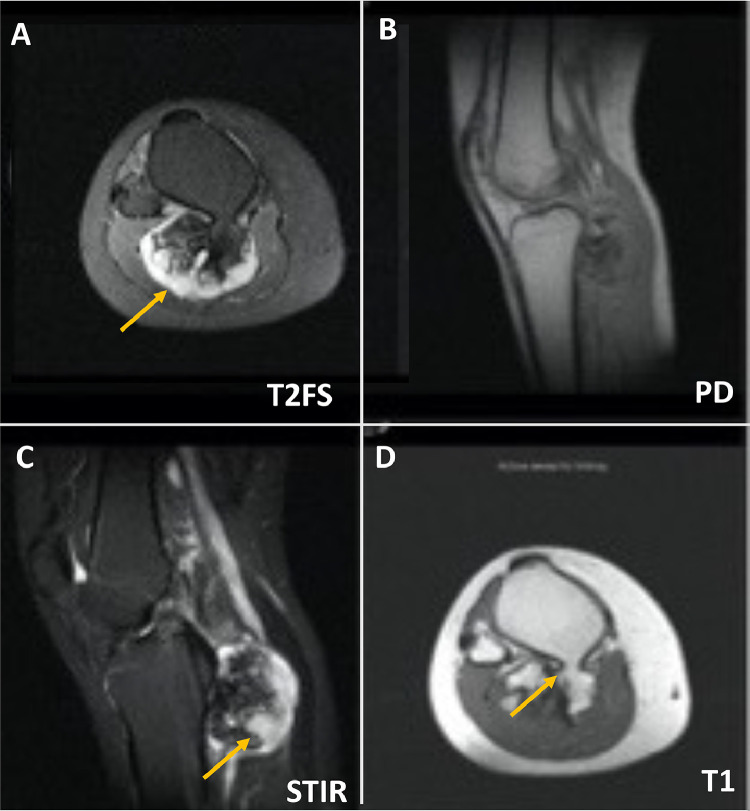
We report a case of tibial osteochondroma in a 25-year-old female who presented with a palpable calf mass. This mass was associated with a thick cartilaginous cap on cross-sectional imaging, suggesting chondrosarcoma. A CT-guided biopsy was performed, and histology, however, was consistent with osteochondroma. Orthopedic oncology recommended surgical excision due to the potential high sampling error with chondroid lesions. The patient underwent surgical resection, resulting in a final diagnosis of osteochondroma. No post-surgical complications occurred, and a 12-month follow-up showed no evidence of local recurrence. This case highlights the atypical imaging feature of a thick cartilaginous cap in a benign etiology without malignant transformation.
我们报告一例25岁女性的胫骨骨软骨瘤病例,该患者小腿有可触及的肿块。在横断面成像中,此肿块伴有厚软骨帽,提示软骨肉瘤。于是进行了CT引导下活检,然而组织学检查结果与骨软骨瘤一致。由于软骨样病变存在潜在的高采样误差,骨肿瘤整形外科建议手术切除。患者接受了手术切除,最终诊断为骨软骨瘤。术后未发生并发症,12个月的随访显示无局部复发迹象。该病例突出了良性病因中厚软骨帽这一非典型影像学特征,且未发生恶性转化。