Raghubeer Nishen, Lahri Sa'ad, Hendrikse Clint
Division of Emergency Medicine, University of Cape Town, Cape Town, South Africa.
Division of Emergency Medicine, University of Stellenbosch, Stellenbosch, South Africa.
Afr J Emerg Med. 2024 Jun;14(2):65-69. doi: 10.1016/j.afjem.2024.01.004. Epub 2024 Feb 21.
Pulmonary embolism (PE) is a significant global cause of mortality, ranking third after myocardial infarction and stroke. ECG findings may play a valuable role in the prognostication of patients with PE, with various ECG abnormalities proving to be reasonable predictors of haemodynamic decompensation, cardiogenic shock, and even mortality. This study aims to assess the value of electrocardiography in predicting inpatient mortality in patients with acute pulmonary embolism, as diagnosed with computed tomography pulmonary angiogram.
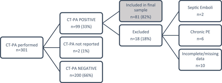
This study was a cross sectional analysis based at Tygerberg Hospital, Cape Town, South Africa. Eligible patients were identified from all CT-PA performed between 1 January 2017 and 31 December 2019 (2 years). The ECGs were independently screened by two blinded emergency physicians for predetermined signs that are associated with right heart strain and higher pulmonary artery pressures, and these findings were analysed to in-hospital mortality.
Of the included 81 patients, 61 (75 %) were female. Of the 41 (51 %) patients with submassive PE and 8 (10 %) with massive PE, 7 (17 %) and 3 (38 %) suffered inpatient mortality ( = 0.023) respectively. Univariate ECG analysis revealed that complete right bundle branch block (OR, 8.6; 95 % CI, 1.1 to 69.9; = 0.044) and right axis deviation (OR, 5.6; 95 % CI, 1.4 to 22.4; = 0.015) were significant predictors of inpatient mortality.
Early identification of patients with pulmonary embolism at higher risk of clinical deterioration and in-patient mortality remains a challenge. Even though no clinical finding or prediction tool in isolation can reliably predict outcomes in patients with pulmonary embolism, this study demonstrated two ECG findings at presentation that were associated with a higher likelihood of inpatient mortality. This single-centre observational study with a small sample precludes concrete conclusions and a large follow-up multi-centre study is advised.
肺栓塞(PE)是全球范围内导致死亡的重要原因,在死因中仅次于心肌梗死和中风,位列第三。心电图表现可能在肺栓塞患者的预后评估中发挥重要作用,各种心电图异常被证明是血流动力学失代偿、心源性休克甚至死亡的合理预测指标。本研究旨在评估心电图在预测经计算机断层扫描肺动脉造影确诊的急性肺栓塞患者住院死亡率方面的价值。
本研究是在南非开普敦泰格堡医院进行的横断面分析。从2017年1月1日至2019年12月31日(2年)期间进行的所有CT-PA检查中确定符合条件的患者。心电图由两名不知情的急诊医生独立筛查,以寻找与右心劳损和较高肺动脉压相关的预定体征,并对这些结果进行分析以评估住院死亡率。
纳入的81例患者中,61例(75%)为女性。在41例(51%)次大面积肺栓塞患者和8例(10%)大面积肺栓塞患者中,分别有7例(17%)和3例(38%)发生住院死亡(P = 0.023)。单因素心电图分析显示,完全性右束支传导阻滞(OR,8.6;95%CI,1.1至69.9;P = 0.044)和电轴右偏(OR,5.6;95%CI,1.4至22.4;P = 0.015)是住院死亡率的重要预测指标。
早期识别具有临床恶化和住院死亡高风险的肺栓塞患者仍然是一项挑战。尽管没有单一的临床发现或预测工具能够可靠地预测肺栓塞患者的预后,但本研究显示了两个与住院死亡率较高可能性相关的心电图表现。这项小样本的单中心观察性研究无法得出确切结论,建议进行大规模的多中心随访研究。