Department of Radiology, Peking University Third Hospital, Beijing, China.
Department of Integration of Chinese and Western Medicine, School of Basic Medical Sciences, Peking University, Beijin, China.
J Imaging Inform Med. 2024 Aug;37(4):1960-1968. doi: 10.1007/s10278-024-01016-x. Epub 2024 Mar 1.
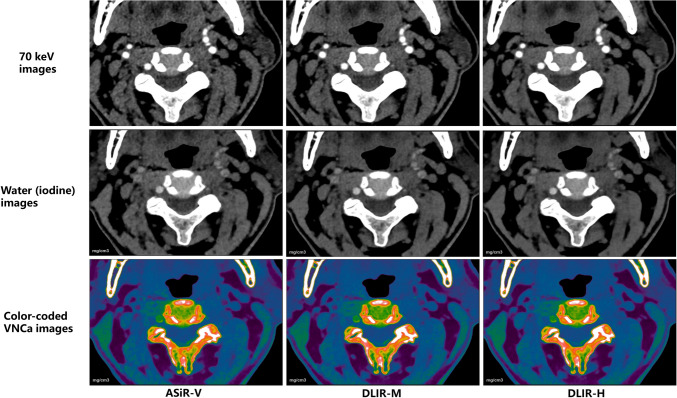
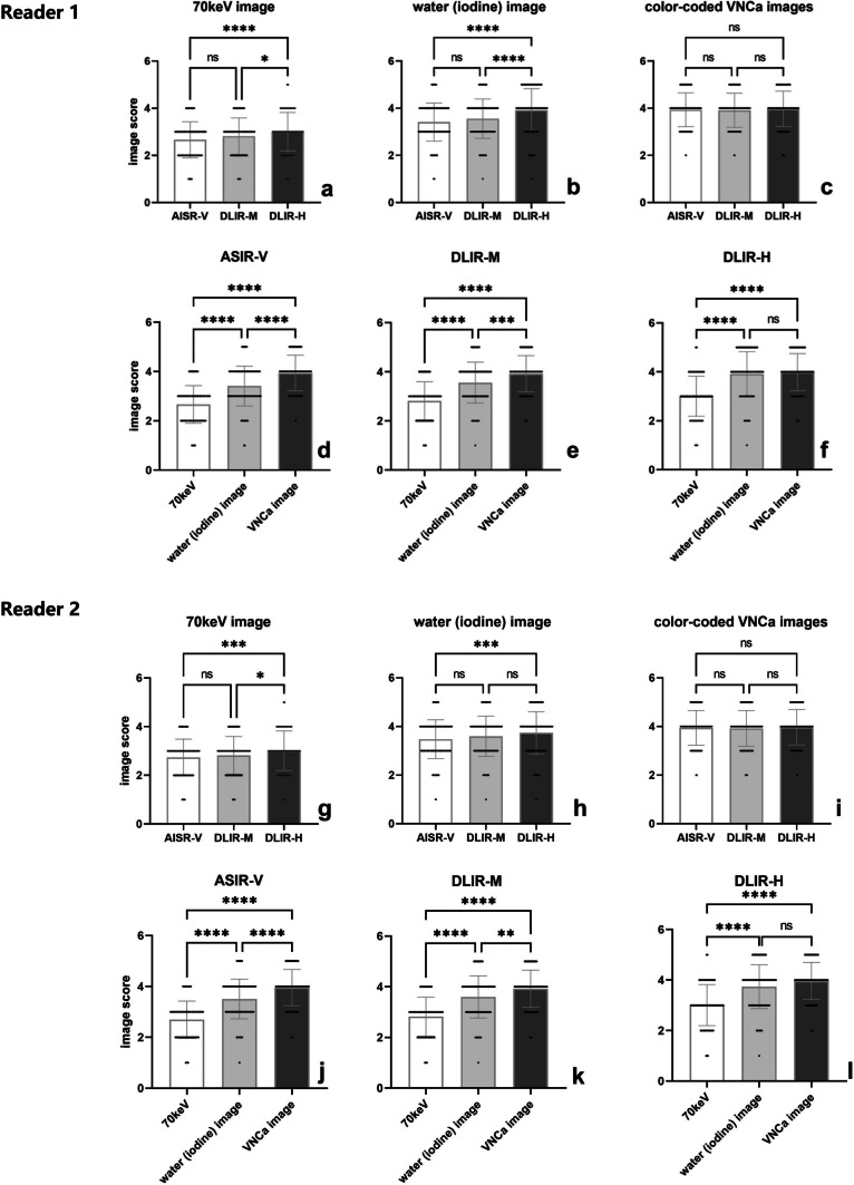
Thus, the aim of this study is to evaluate the performance of deep learning imaging reconstruction (DLIR) algorithm in different image sets derived from carotid dual-energy computed tomography angiography (DECTA) for evaluating cervical intervertebral discs (IVDs) and compare them with those reconstructed using adaptive statistical iterative reconstruction-Veo (ASiR-V). Forty-two patients who underwent carotid DECTA were included in this retrospective analysis. Three types of image sets (70 keV, water-iodine, and water-calcium) were reconstructed using 50% ASiR-V and DLIR at medium and high levels (DLIR-M and DLIR-H). The diagnostic acceptability and conspicuity of IVDs were assessed using a 5-point scale. Hounsfield Units (HU) and water concentration (WC) values of the IVDs; standard deviation (SD); and coefficient of variation (CV) were calculated. Measurement parameters of the 50% ASIR-V, DLIR-M, and DLIR-H groups were compared. The DLIR-H group showed higher scores for diagnostic acceptability and conspicuity, as well as lower SD values for HU and WC than the ASiR-V and DLIR-M groups for the 70 keV and water-iodine image sets (all p < .001). However, there was no significant difference in scores and SD among the three groups for the water-calcium image set (all p > .005). The water-calcium image set showed better diagnostic accuracy for evaluating IVDs compared to the other image sets. The inter-rater agreement using ASiR-V, DLIR-M, and DLIR-H was good for the 70 keV image set, excellent for the water-iodine and water-calcium image sets. DLIR improved the visualization of IVDs in the 70 keV and water-iodine image sets. However, its improvement on color-coded water-calcium image set was limited.
因此,本研究旨在评估深度学习成像重建(DLIR)算法在颈动脉双能 CT 血管造影(DECTA)不同图像组中的性能,用于评估颈椎间盘(IVD),并将其与自适应统计迭代重建-Veo(ASiR-V)重建的图像进行比较。本回顾性分析纳入了 42 例接受颈动脉 DECTA 检查的患者。使用 50%ASiR-V 和中、高级 DLIR(DLIR-M 和 DLIR-H)重建 70keV、水碘和水钙三种图像组。使用 5 分制评估 IVD 的诊断可接受性和显影性。计算 IVD 的 Hounsfield 单位(HU)和水浓度(WC)值、标准差(SD)和变异系数(CV)。比较 50%ASiR-V、DLIR-M 和 DLIR-H 组的测量参数。与 ASiR-V 和 DLIR-M 组相比,DLIR-H 组在 70keV 和水碘图像组中,诊断可接受性和显影性评分较高,HU 和 WC 的 SD 值较低(均 p<0.001)。然而,在水钙图像组中,三组间评分和 SD 无显著差异(均 p>0.005)。与其他图像组相比,水钙图像组对 IVD 的评估具有更好的诊断准确性。使用 ASiR-V、DLIR-M 和 DLIR-H 的组内一致性在 70keV 图像组中良好,在水碘和水钙图像组中优秀。DLIR 改善了 70keV 和水碘图像组中 IVD 的可视化效果。然而,其对彩色编码水钙图像组的改善效果有限。