Kraus Abigayle C, Iqbal Zohaib, Cardan Rex A, Popple Richard A, Stanley Dennis N, Shen Sui, Pogue Joel A, Wu Xingen, Lee Kevin, Marcrom Samuel, Cardenas Carlos E
Heersink School of Medicine, University of Alabama at Birmingham, Birmingham, Alabama.
Department of Radiation Oncology, University of Alabama at Birmingham, Birmingham, Alabama.
Adv Radiat Oncol. 2023 Dec 10;9(4):101417. doi: 10.1016/j.adro.2023.101417. eCollection 2024 Apr.
The use of deep learning to auto-contour organs at risk (OARs) in gynecologic radiation treatment is well established. Yet, there is limited data investigating the prospective use of auto-contouring in clinical practice. In this study, we assess the accuracy and efficiency of auto-contouring OARs for computed tomography-based brachytherapy treatment planning of gynecologic malignancies.
An inhouse contouring tool automatically delineated 5 OARs in gynecologic radiation treatment planning: the bladder, small bowel, sigmoid, rectum, and urethra. Accuracy of each auto-contour was evaluated using a 5-point Likert scale: a score of 5 indicated the contour could be used without edits, while a score of 1 indicated the contour was unusable. During scoring, automated contours were edited and subsequently used for treatment planning. Dice similarity coefficient, mean surface distance, 95% Hausdorff distance, Hausdorff distance, and dosimetric changes between original and edited contours were calculated. Contour approval time and total planning time of a prospective auto-contoured (AC) cohort were compared with times from a retrospective manually contoured (MC) cohort.
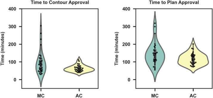
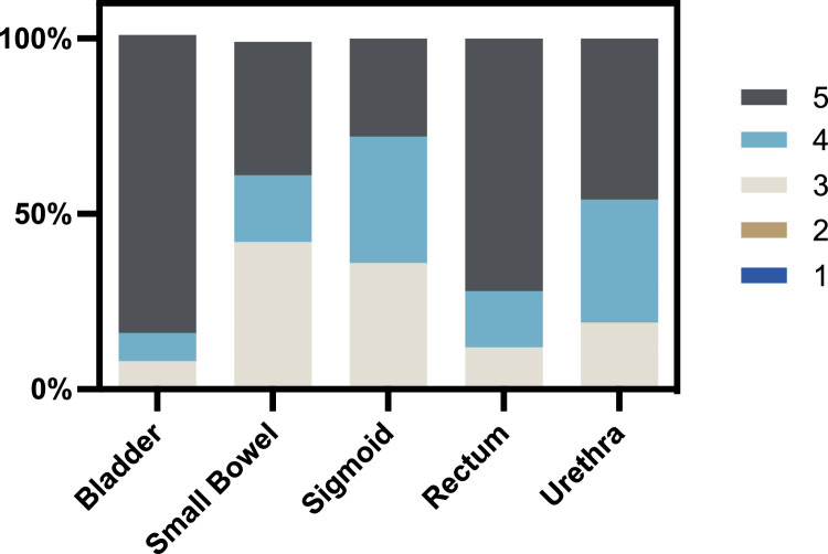
Thirty AC cases from January 2022 to July 2022 and 31 MC cases from July 2021 to January 2022 were included. The mean (±SD) Likert score for each OAR was the following: bladder 4.77 (±0.58), small bowel 3.96 (±0.91), sigmoid colon 3.92 (±0.81), rectum 4.6 (±0.71), and urethra 4.27 (±0.78). No ACs required major edits. All OARs had a mean Dice similarity coefficient > 0.86, mean surface distance < 0.48 mm, 95% Hausdorff distance < 3.2 mm, and Hausdorff distance < 10.32 mm between original and edited contours. There was no significant difference in dose-volume histogram metrics (D2.0 cc/D0.1 cc) between original and edited contours ( values > .05). The average time to plan approval in the AC cohort was 19% less than the MC cohort. (AC vs MC, 117.0 + 18.0 minutes vs 144.9 ± 64.5 minutes, = .045).
Automated contouring is useful and accurate in clinical practice. Auto-contouring OARs streamlines radiation treatment workflows and decreases time required to design and approve gynecologic brachytherapy plans.
深度学习用于妇科放射治疗中危及器官(OARs)的自动轮廓勾画已得到广泛应用。然而,关于自动轮廓勾画在临床实践中的前瞻性应用的数据有限。在本研究中,我们评估了基于计算机断层扫描的近距离放射治疗计划中,妇科恶性肿瘤OARs自动轮廓勾画的准确性和效率。
一个内部轮廓勾画工具在妇科放射治疗计划中自动勾勒出5个OARs:膀胱、小肠、乙状结肠、直肠和尿道。使用5点李克特量表评估每个自动轮廓的准确性:5分表示轮廓无需编辑即可使用,1分表示轮廓不可用。在评分过程中,对自动轮廓进行编辑,随后用于治疗计划。计算原始轮廓和编辑后轮廓之间的骰子相似系数、平均表面距离、95%豪斯多夫距离、豪斯多夫距离和剂量学变化。将前瞻性自动轮廓勾画(AC)队列的轮廓批准时间和总计划时间与回顾性手动轮廓勾画(MC)队列的时间进行比较。
纳入了2022年1月至2022年7月的30例AC病例和2021年7月至2022年1月的31例MC病例。每个OAR的平均(±标准差)李克特评分为:膀胱4.77(±0.58),小肠3.96(±0.91),乙状结肠3.92(±0.81),直肠4.6(±0.71),尿道4.27(±0.78)。没有AC需要进行重大编辑。所有OARs在原始轮廓和编辑后轮廓之间的平均骰子相似系数>0.86,平均表面距离<0.48毫米,95%豪斯多夫距离<3.2毫米,豪斯多夫距离<10.32毫米。原始轮廓和编辑后轮廓之间的剂量体积直方图指标(D2.0 cc/D0.1 cc)没有显著差异(P值>.05)。AC队列中计划批准的平均时间比MC队列少19%。(AC与MC,117.0 + 18.0分钟对144.9 ± 64.5分钟,P =.045)。
自动轮廓勾画在临床实践中是有用且准确的。OARs的自动轮廓勾画简化了放射治疗工作流程,并减少了设计和批准妇科近距离放射治疗计划所需的时间。