Bethune Third Clinical Medical College, Jilin University, Changchun, 130021, China.
Department of Gastrointestinal, Colorectal and Anal Surgery, China-Japan Union Hospital of Jilin University, NO. 126, Xiantai Street, Changchun, 130033, China.
Cell Oncol (Dordr). 2024 Aug;47(4):1113-1126. doi: 10.1007/s13402-024-00935-9. Epub 2024 Mar 23.
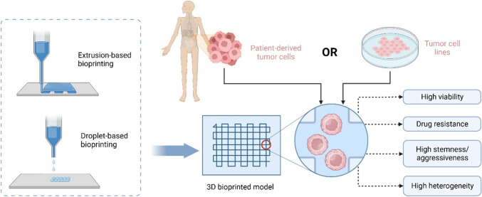
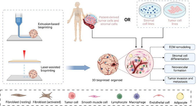
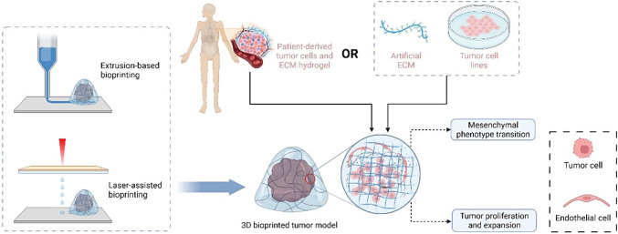
Cancer immunotherapy is receiving worldwide attention for its induction of an anti-tumor response. However, it has had limited efficacy in some patients who acquired resistance. The dynamic and sophisticated complexity of the tumor microenvironment (TME) is the leading contributor to this clinical dilemma. Through recapitulating the physiological features of the TME, 3D bioprinting is a promising research tool for cancer immunotherapy, which preserves in vivo malignant aggressiveness, heterogeneity, and the cell-cell/matrix interactions. It has been reported that application of 3D bioprinting holds potential to address the challenges of immunotherapy resistance and facilitate personalized medication.
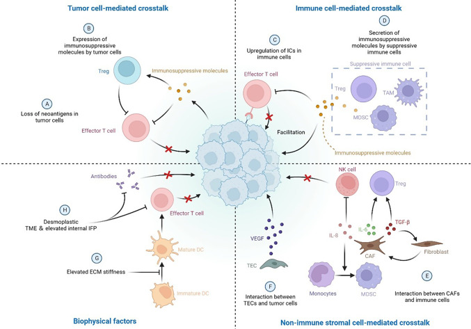
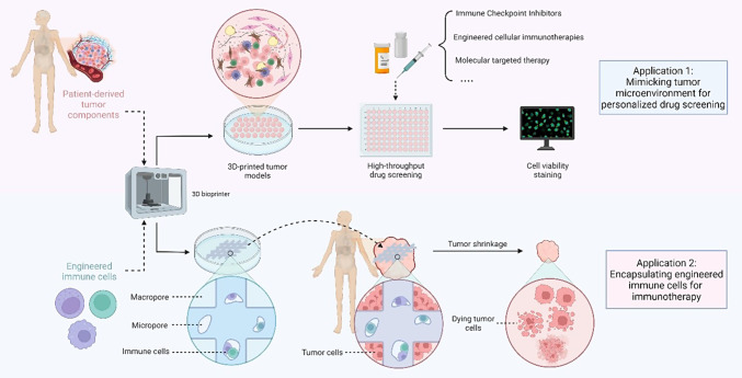
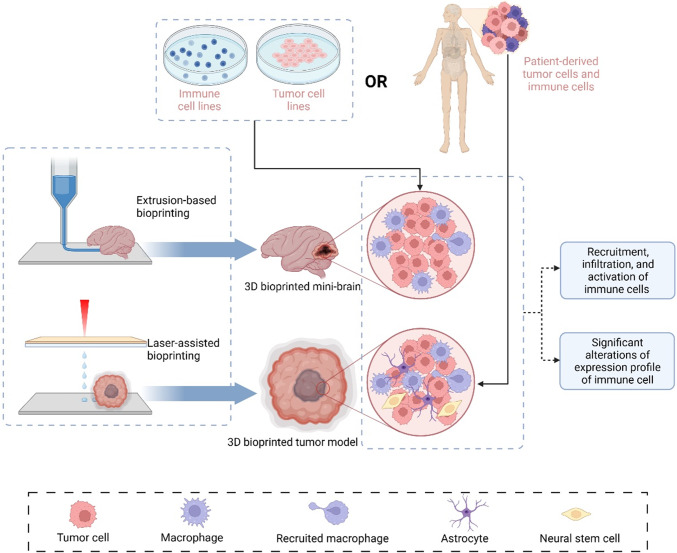
In this review, we briefly summarize the contributions of cellular and noncellular components of the TME in the development of immunotherapy resistance, and introduce recent advances in 3D bioprinted tumor models that served as platforms to study the interactions between tumor cells and the TME. By constructing multicellular 3D bioprinted tumor models, cellular and noncellular crosstalk is reproduced between tumor cells, immune cells, fibroblasts, adipocytes, and the extracellular matrix (ECM) within the TME. In the future, by quickly preparing 3D bioprinted tumor models with patient-derived components, information on tumor immunotherapy resistance can be obtained timely for clinical reference. The combined application with tumoroid or other 3D culture technologies will also help to better simulate the complexity and dynamics of tumor microenvironment in vitro. We aim to provide new perspectives for overcoming cancer immunotherapy resistance and inspire multidisciplinary research to improve the clinical application of 3D bioprinting technology.
癌症免疫疗法因其诱导抗肿瘤反应而受到全球关注。然而,在一些产生耐药性的患者中,其疗效有限。肿瘤微环境(TME)的动态和复杂复杂性是导致这一临床困境的主要原因。通过再现 TME 的生理特征,3D 生物打印是癌症免疫疗法的一种有前途的研究工具,它保留了体内恶性侵袭性、异质性和细胞-细胞/基质相互作用。据报道,应用 3D 生物打印有可能解决免疫疗法耐药性的挑战,并促进个性化药物治疗。
在这篇综述中,我们简要总结了 TME 的细胞和非细胞成分在免疫疗法耐药性发展中的作用,并介绍了 3D 生物打印肿瘤模型的最新进展,这些模型可作为研究肿瘤细胞与 TME 之间相互作用的平台。通过构建多细胞 3D 生物打印肿瘤模型,可以再现 TME 中肿瘤细胞、免疫细胞、成纤维细胞、脂肪细胞和细胞外基质(ECM)之间的细胞和非细胞串扰。在未来,通过快速制备具有患者来源成分的 3D 生物打印肿瘤模型,可以及时获得有关肿瘤免疫疗法耐药性的信息,为临床参考。与肿瘤球或其他 3D 培养技术的联合应用也将有助于更好地模拟肿瘤微环境的复杂性和动态性。我们旨在为克服癌症免疫疗法耐药性提供新的视角,并激发多学科研究,以改善 3D 生物打印技术的临床应用。