Liu Mengyi, Ji Zhicheng, Jain Vaibhav, Smith Vanessa L, Hocke Emily, Patel Anoop P, McLendon Roger E, Ashley David M, Gregory Simon G, López Giselle Y
Computational Biology and Bioinformatics Program, Duke University School of Medicine, Durham, NC, 27710, USA.
The Preston Robert Tisch Brain Tumor Center, Duke University School of Medicine, Durham, NC, 27710, USA.
Acta Neuropathol Commun. 2024 Apr 22;12(1):64. doi: 10.1186/s40478-024-01769-0.
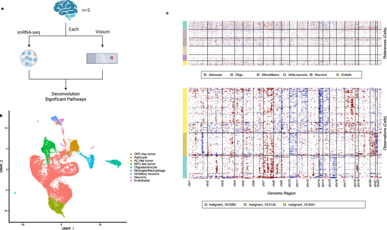
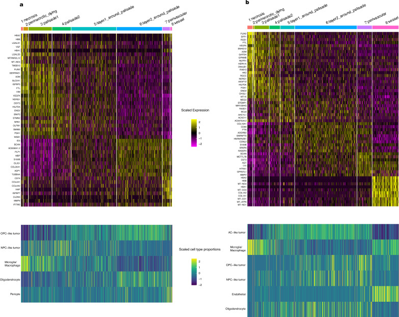
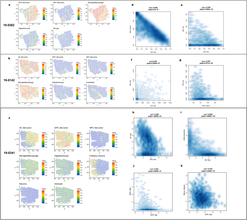
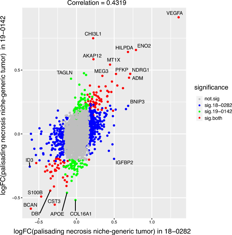
Glioblastoma (GBM) remains an untreatable malignant tumor with poor patient outcomes, characterized by palisading necrosis and microvascular proliferation. While single-cell technology made it possible to characterize different lineage of glioma cells into neural progenitor-like (NPC-like), oligodendrocyte-progenitor-like (OPC-like), astrocyte-like (AC-like) and mesenchymal like (MES-like) states, it does not capture the spatial localization of these tumor cell states. Spatial transcriptomics empowers the study of the spatial organization of different cell types and tumor cell states and allows for the selection of regions of interest to investigate region-specific and cell-type-specific pathways. Here, we obtained paired 10x Chromium single-nuclei RNA-sequencing (snRNA-seq) and 10x Visium spatial transcriptomics data from three GBM patients to interrogate the GBM microenvironment. Integration of the snRNA-seq and spatial transcriptomics data reveals patterns of segregation of tumor cell states. For instance, OPC-like tumor and NPC-like tumor significantly segregate in two of the three samples. Our differentially expressed gene and pathway analyses uncovered significant pathways in functionally relevant niches. Specifically, perinecrotic regions were more immunosuppressive than the endogenous GBM microenvironment, and perivascular regions were more pro-inflammatory. Our gradient analysis suggests that OPC-like tumor cells tend to reside in areas closer to the tumor vasculature compared to tumor necrosis, which may reflect increased oxygen requirements for OPC-like cells. In summary, we characterized the localization of cell types and tumor cell states, the gene expression patterns, and pathways in different niches within the GBM microenvironment. Our results provide further evidence of the segregation of tumor cell states and highlight the immunosuppressive nature of the necrotic and perinecrotic niches in GBM.
胶质母细胞瘤(GBM)仍然是一种无法治愈的恶性肿瘤,患者预后较差,其特征为栅栏状坏死和微血管增殖。虽然单细胞技术能够将不同谱系的胶质瘤细胞表征为神经祖细胞样(NPC样)、少突胶质细胞祖细胞样(OPC样)、星形胶质细胞样(AC样)和间充质样(MES样)状态,但它无法捕捉这些肿瘤细胞状态的空间定位。空间转录组学有助于研究不同细胞类型和肿瘤细胞状态的空间组织,并允许选择感兴趣的区域来研究区域特异性和细胞类型特异性途径。在这里,我们从三名GBM患者中获得了配对的10x铬单细胞RNA测序(snRNA-seq)和10x Visium空间转录组学数据,以探究GBM微环境。snRNA-seq和空间转录组学数据的整合揭示了肿瘤细胞状态的分离模式。例如,在三个样本中的两个样本中,OPC样肿瘤和NPC样肿瘤显著分离。我们的差异表达基因和通路分析揭示了功能相关微环境中的重要通路。具体而言,坏死周围区域比内源性GBM微环境更具免疫抑制性,而血管周围区域更具促炎性。我们的梯度分析表明,与肿瘤坏死相比,OPC样肿瘤细胞倾向于驻留在更靠近肿瘤血管的区域,这可能反映了OPC样细胞对氧气需求的增加。总之,我们表征了GBM微环境中不同微环境内的细胞类型和肿瘤细胞状态的定位、基因表达模式和通路。我们的结果为肿瘤细胞状态的分离提供了进一步的证据,并突出了GBM中坏死和坏死周围微环境的免疫抑制性质。