Kalmegh Padmashri P, Hande Alka
Department of Oral and Maxillofacial Pathology, Sharad Pawar Dental College and Hospital, Datta Meghe Institute of Higher Education and Research, Wardha, IND.
Department of Oral Pathology and Microbiology, Sharad Pawar Dental College and Hospital, Datta Meghe Institute of Higher Education and Research, Wardha, IND.
Cureus. 2024 Mar 23;16(3):e56771. doi: 10.7759/cureus.56771. eCollection 2024 Mar.
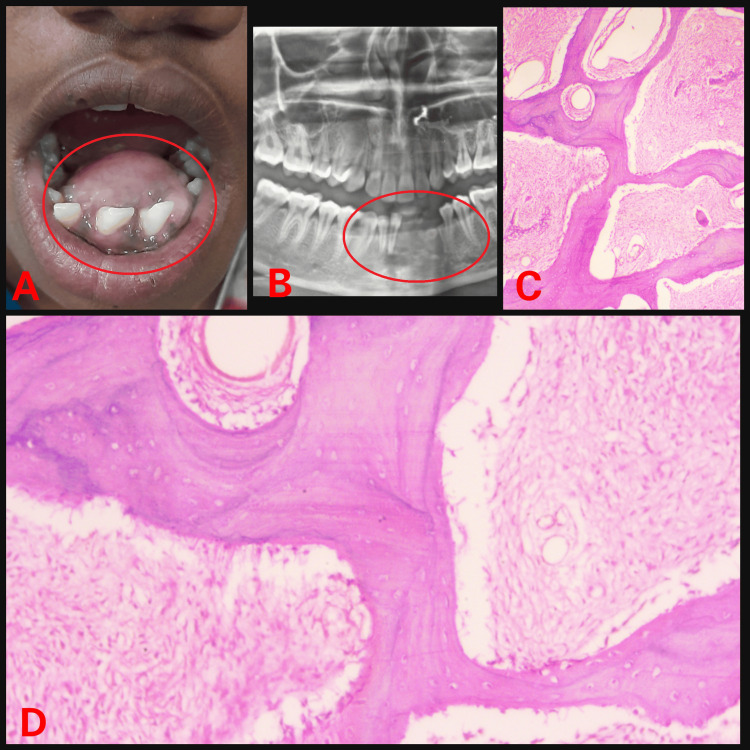
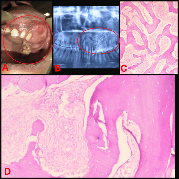
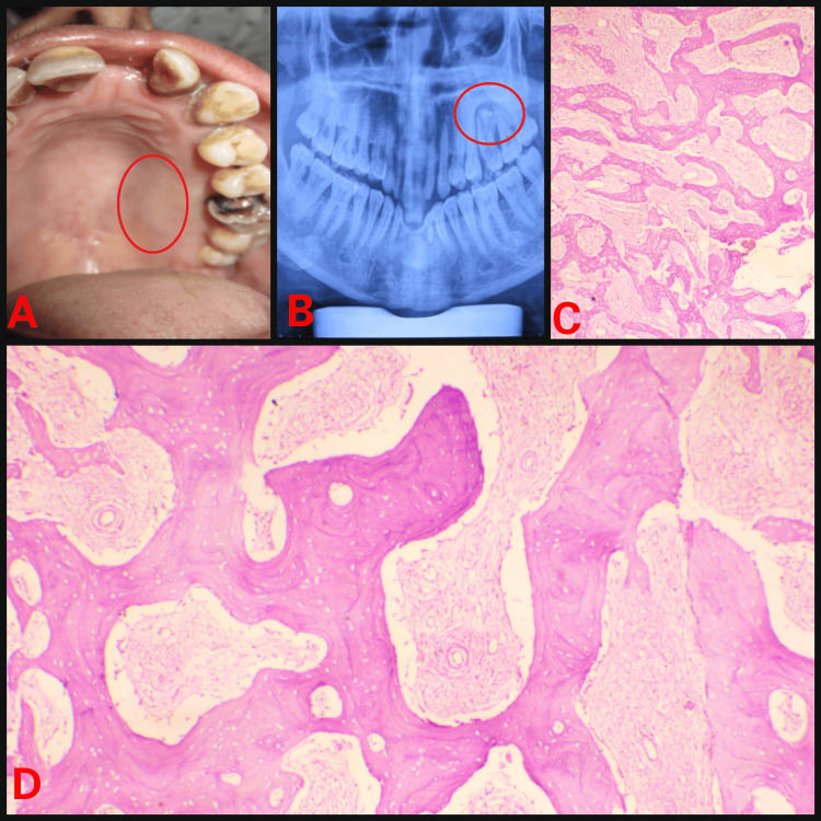
Craniofacial fibro-osseous lesions (CFOLs) are a diverse group of relatively rare entities whose etiology ranges from reactive to dysplastic with a potential for malignant transformation. It is distinguished by the replacement of bone with fibrous tissue, that subsequently develops different degrees of calcification. Fibrous dysplasia (FD) is a component of the fibro-osseous lesion spectrum. The clinical spectrum of FD is wide, ranging from minor monostotic lesions affecting a single bone to devastating polyostotic disease involving the entire skeleton. FD produces asymmetry, which impairs face aesthetics. FD leads to bone differentiation, disintegration, and disorganization. It depicts a cellular collagenous stroma lacking mitotic figures and pleomorphism. Blood capillaries are evenly distributed, as are elongated trabeculae of woven or lamellar bone with uneven curves (often referred to as the Chinese letters pattern). Three types of FD patterns can be identified by computed tomography (CT) imaging: a cystic pattern, a homogeneously dense pattern, and a ground-glass pattern. The cornerstone of treatment is surgery, although the method varies depending on the location, size, and symptoms of the lesion. As an alternative to surgery, the use of bisphosphonates to reduce osteoclastic activity is under consideration. In this case series, we present three cases of FD involving the maxilla and mandible. We aim to correlate the clinical presentation, histological features, and radiographic findings, to promote early diagnosis, treatment, and better prognosis of the patient.
颅面纤维性骨病变(CFOLs)是一组多样的相对罕见的病症,其病因从反应性到发育异常不等,具有恶变潜能。其特征是骨被纤维组织替代,随后发展出不同程度的钙化。纤维发育不良(FD)是纤维性骨病变谱的一个组成部分。FD的临床谱很广,从影响单一骨骼的轻度单骨病变到累及整个骨骼的严重多骨病变。FD会导致不对称,损害面部美观。FD会导致骨分化、崩解和结构紊乱。它表现为缺乏有丝分裂象和多形性的细胞胶原基质。毛细血管均匀分布,编织骨或板层骨的细长小梁也均匀分布,其曲线不均匀(常称为“中国字母”图案)。通过计算机断层扫描(CT)成像可识别出三种FD模式:囊性模式、均匀致密模式和磨玻璃模式。治疗的基石是手术,不过方法会根据病变的位置、大小和症状而有所不同。作为手术的替代方法,正在考虑使用双膦酸盐来降低破骨细胞活性。在本病例系列中,我们展示了三例累及上颌骨和下颌骨的FD病例。我们旨在将临床表现、组织学特征和影像学表现相关联,以促进患者的早期诊断、治疗和更好预后。