Raventos-Suarez C, Kaul D K, Macaluso F, Nagel R L
Proc Natl Acad Sci U S A. 1985 Jun;82(11):3829-33. doi: 10.1073/pnas.82.11.3829.
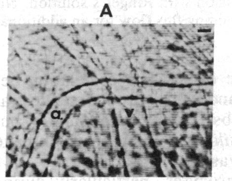
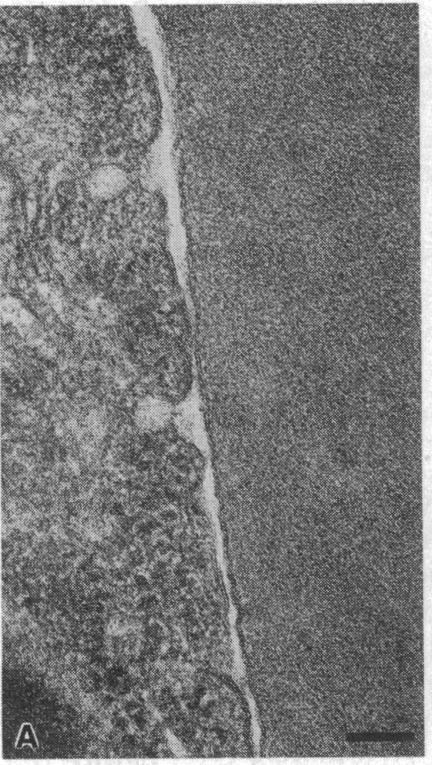
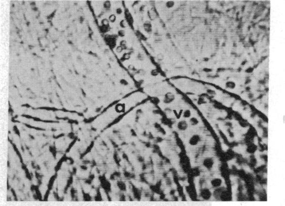
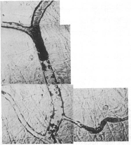
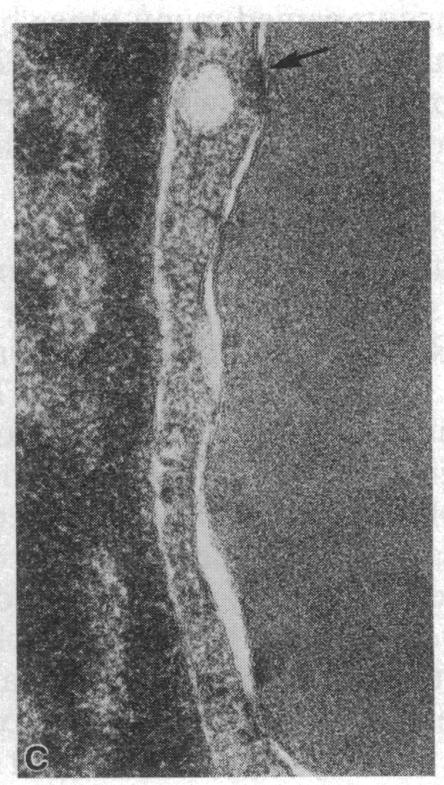
We have studied the pathophysiology of the vascular obstruction induced by Plasmodium falciparum-parasitized erythrocytes with the use of an ex vivo microcirculatory preparation perfused with red cells infected with knobless and knobby clones of the FCR-3 strain. We find that parasitized erythrocyte membrane knobs are indispensable for the generation of the circulatory obstruction. Uninfected erythrocytes incubated in culture and erythrocytes infected with early or late forms of the knobless clones or the early forms of the knobby clone all failed to obstruct the microcirculation, although exhibiting various effects on bulk viscosity and peripheral resistance during flow. In contrast, late forms of the knobby clone produced significantly higher peripheral resistance during flow and significant obstruction as detected by changes in time of pressure flow recovery as well as by direct videorecorded microscopic observation. Optical and electron microscopy showed that the adherence of parasitized cells to the endothelium was limited to the venules and involved the knobs in junctions. In addition, we were able to follow the sequence of events during obstruction: initial red-cell adherence to the venular endothelium (sometimes only transitory) followed by progressive recruitment at the venule surface, finally leading to total obstruction that involved parasitized and nonparasitized erythrocytes. Sometimes, retrograde aggregation would extend the obstruction to the capillaries or even precapillary arterioles. These results show that knobs are necessary and sufficient to produce vascular obstruction and that other factors (spleen, immunological, etc.) can only have a modulating role. These results also exclude the possibility that the exclusive adherence to venules is the consequence of "plasma factors" found in the malaric patients.
我们利用一种体外微循环制剂,灌注感染了FCR - 3株无结和有结克隆的红细胞,研究了恶性疟原虫寄生红细胞诱导的血管阻塞的病理生理学。我们发现,寄生红细胞膜上的结对于循环阻塞的产生是必不可少的。在培养中孵育的未感染红细胞以及感染无结克隆早期或晚期形式或有结克隆早期形式的红细胞,尽管在流动过程中对总体粘度和外周阻力表现出各种影响,但都未能阻塞微循环。相比之下,有结克隆的晚期形式在流动过程中产生了显著更高的外周阻力,并且通过压力流恢复时间的变化以及直接的视频记录显微镜观察检测到显著的阻塞。光学和电子显微镜显示,寄生细胞与内皮的粘附仅限于小静脉,并且在连接处涉及到结。此外,我们能够追踪阻塞过程中的一系列事件:最初红细胞粘附于小静脉内皮(有时只是短暂的),随后在小静脉表面逐渐募集,最终导致包括寄生和未寄生红细胞的完全阻塞。有时,逆行聚集会将阻塞扩展到毛细血管甚至毛细血管前小动脉。这些结果表明,结对于产生血管阻塞是必要且充分的,而其他因素(脾脏、免疫等)只能起到调节作用。这些结果也排除了仅粘附于小静脉是疟疾患者中发现的“血浆因子”所致的可能性。