Kanan Achraf, Pereira Bruno, Hordonneau Constance, Cassagnes Lucie, Pouget Eléonore, Tianhoun Léon Appolinaire, Chauveau Benoît, Magnin Benoît
Department of Radiology, Estaing Hospital, Clermont University Hospital, Clermont-Ferrand, France.
Department of Biostatistics, DRCI, Clermont University Hospital, Clermont-Ferrand, France.
Insights Imaging. 2024 Jul 6;15(1):167. doi: 10.1186/s13244-024-01753-1.
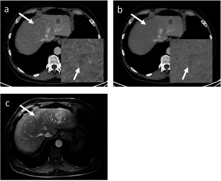
Detection of liver metastases is crucial for guiding oncological management. Computed tomography through iterative reconstructions is widely used in this indication but has certain limitations. Deep learning image reconstructions (DLIR) use deep neural networks to achieve a significant noise reduction compared to iterative reconstructions. While reports have demonstrated improvements in image quality, their impact on liver metastases detection remains unclear. Our main objective was to determine whether DLIR affects the number of detected liver metastasis. Our secondary objective was to compare metastases conspicuity between the two reconstruction methods.
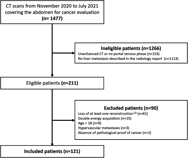
CT images of 121 patients with liver metastases were reconstructed using a 50% adaptive statistical iterative reconstruction (50%-ASiR-V), and three levels of DLIR (DLIR-low, DLIR-medium, and DLIR-high). For each reconstruction, two double-blinded radiologists counted up to a maximum of ten metastases. Visibility and contour definitions were also assessed. Comparisons between methods for continuous parameters were performed using mixed models.
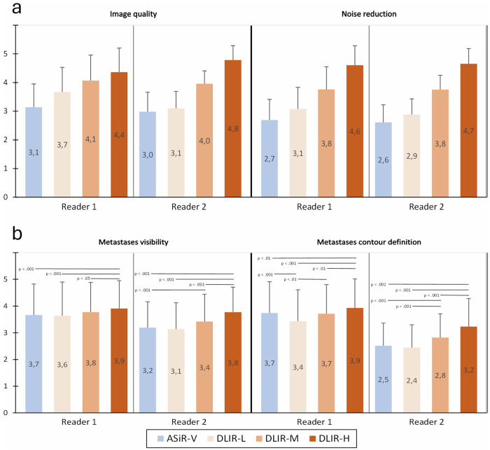
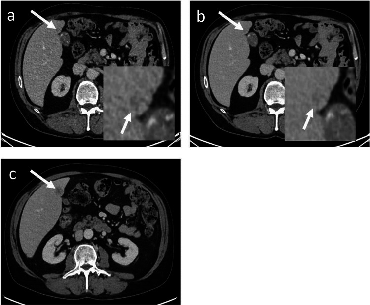
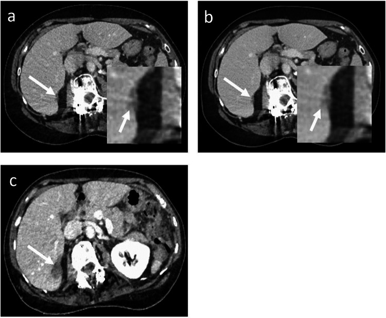
A higher number of metastases was detected by one reader with DLIR-high: 7 (2-10) (median (Q₁-Q₃); total 733) versus 5 (2-10), respectively for DLIR-medium, DLIR-low, and ASiR-V (p < 0.001). Ten patents were detected with more metastases with DLIR-high simultaneously by both readers and a third reader for confirmation. Metastases visibility and contour definition were better with DLIR than ASiR-V.
DLIR-high enhanced the detection and visibility of liver metastases compared to ASiR-V, and also increased the number of liver metastases detected.
Deep learning-based reconstruction at high strength allowed an increase in liver metastases detection compared to hybrid iterative reconstruction and can be used in clinical oncology imaging to help overcome the limitations of CT.
Detection of liver metastases is crucial but limited with standard CT reconstructions. More liver metastases were detected with deep-learning CT reconstruction compared to iterative reconstruction. Deep learning reconstructions are suitable for hepatic metastases staging and follow-up.
检测肝转移对于指导肿瘤治疗至关重要。通过迭代重建的计算机断层扫描在该适应症中广泛使用,但有一定局限性。深度学习图像重建(DLIR)使用深度神经网络,与迭代重建相比可显著降低噪声。虽然报告显示图像质量有所改善,但其对肝转移检测的影响仍不明确。我们的主要目的是确定DLIR是否会影响检测到的肝转移数量。次要目的是比较两种重建方法之间转移灶的清晰度。
使用50%自适应统计迭代重建(50%-ASiR-V)以及三种水平的DLIR(低水平DLIR、中等水平DLIR和高水平DLIR)对121例肝转移患者的CT图像进行重建。对于每种重建,两名双盲放射科医生最多计数十个转移灶。还评估了可见性和轮廓清晰度。使用混合模型对连续参数的方法之间进行比较。
一名读者使用高水平DLIR检测到的转移灶数量更多:7(2 - 10)(中位数(Q₁ - Q₃);总计733个),而中等水平DLIR、低水平DLIR和ASiR-V分别为5(2 - 10)(p < 0.001)。两名读者同时使用高水平DLIR检测到有更多转移灶的十名患者,由第三名读者进行确认。与ASiR-V相比,DLIR的转移灶可见性和轮廓清晰度更好。
与ASiR-V相比,高水平DLIR增强了肝转移的检测和可见性,还增加了检测到的肝转移数量。
与混合迭代重建相比,高强度基于深度学习的重建可增加肝转移的检测,可用于临床肿瘤成像以帮助克服CT的局限性。
肝转移的检测至关重要,但标准CT重建存在局限性。与迭代重建相比,深度学习CT重建检测到更多肝转移。深度学习重建适用于肝转移分期和随访。