• 文献检索
  • 文档翻译
  • 深度研究
  • 学术资讯
  • Suppr Zotero 插件Zotero 插件
  • 邀请有礼
  • 套餐&价格
  • 历史记录
应用&插件
Suppr Zotero 插件Zotero 插件浏览器插件Mac 客户端Windows 客户端微信小程序
定价
高级版会员购买积分包购买API积分包
服务
文献检索文档翻译深度研究API 文档MCP 服务
关于我们
关于 Suppr公司介绍联系我们用户协议隐私条款
关注我们

Suppr 超能文献

核心技术专利:CN118964589B侵权必究
粤ICP备2023148730 号-1Suppr @ 2026

文献检索

告别复杂PubMed语法,用中文像聊天一样搜索,搜遍4000万医学文献。AI智能推荐,让科研检索更轻松。

立即免费搜索

文件翻译

保留排版,准确专业,支持PDF/Word/PPT等文件格式,支持 12+语言互译。

免费翻译文档

深度研究

AI帮你快速写综述,25分钟生成高质量综述,智能提取关键信息,辅助科研写作。

立即免费体验

COVLIAS 3.0:基于云的量化混合UNet3+深度学习用于肺部计算机断层扫描中的新冠肺炎病变检测

COVLIAS 3.0: cloud-based quantized hybrid UNet3+ deep learning for COVID-19 lesion detection in lung computed tomography.

作者信息

Agarwal Sushant, Saxena Sanjay, Carriero Alessandro, Chabert Gian Luca, Ravindran Gobinath, Paul Sudip, Laird John R, Garg Deepak, Fatemi Mostafa, Mohanty Lopamudra, Dubey Arun K, Singh Rajesh, Fouda Mostafa M, Singh Narpinder, Naidu Subbaram, Viskovic Klaudija, Kukuljan Melita, Kalra Manudeep K, Saba Luca, Suri Jasjit S

机构信息

Advanced Knowledge Engineering Center, GBTI, Roseville, CA, United States.

Department of CSE, PSIT, Kanpur, India.

出版信息

Front Artif Intell. 2024 Jun 28;7:1304483. doi: 10.3389/frai.2024.1304483. eCollection 2024.

DOI:
10.3389/frai.2024.1304483
PMID:39006802
原文链接:https://pmc.ncbi.nlm.nih.gov/articles/PMC11240867/
Abstract

BACKGROUND AND NOVELTY

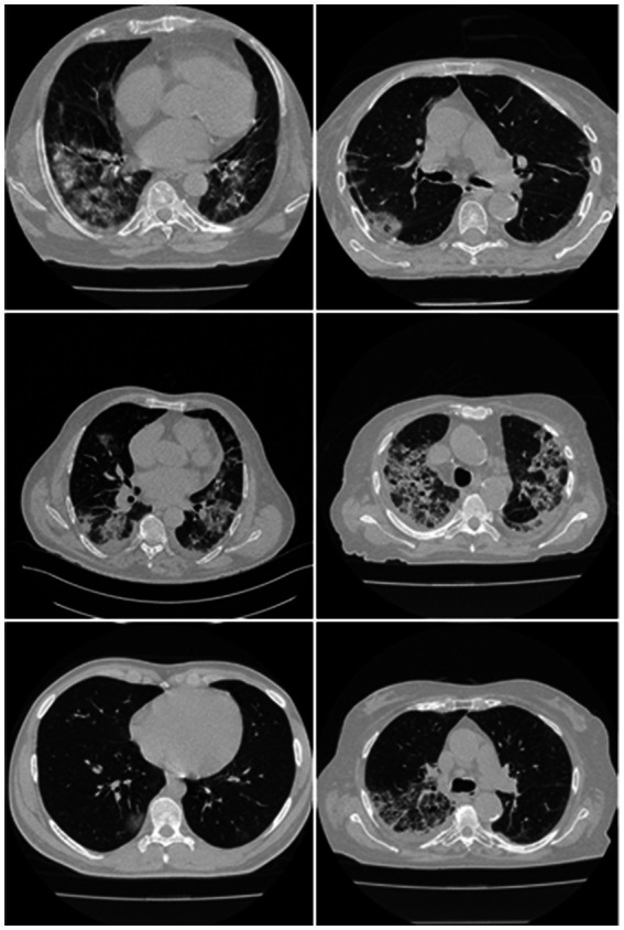
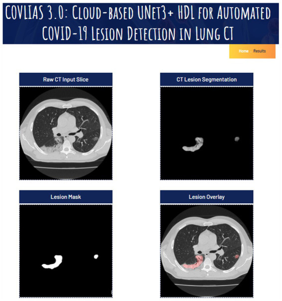
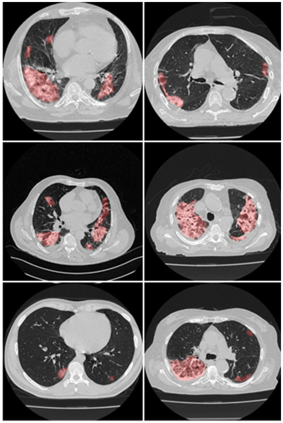
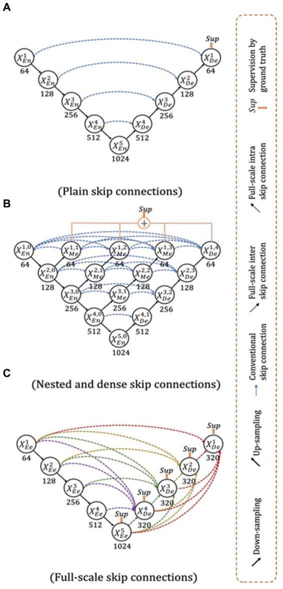
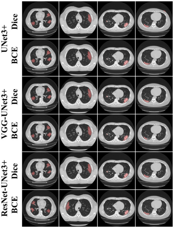
When RT-PCR is ineffective in early diagnosis and understanding of COVID-19 severity, Computed Tomography (CT) scans are needed for COVID diagnosis, especially in patients having high ground-glass opacities, consolidations, and crazy paving. Radiologists find the manual method for lesion detection in CT very challenging and tedious. Previously solo deep learning (SDL) was tried but they had low to moderate-level performance. This study presents two new cloud-based quantized deep learning UNet3+ hybrid (HDL) models, which incorporated full-scale skip connections to enhance and improve the detections.

METHODOLOGY

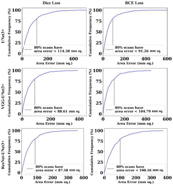
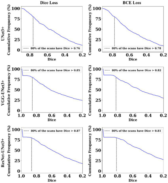
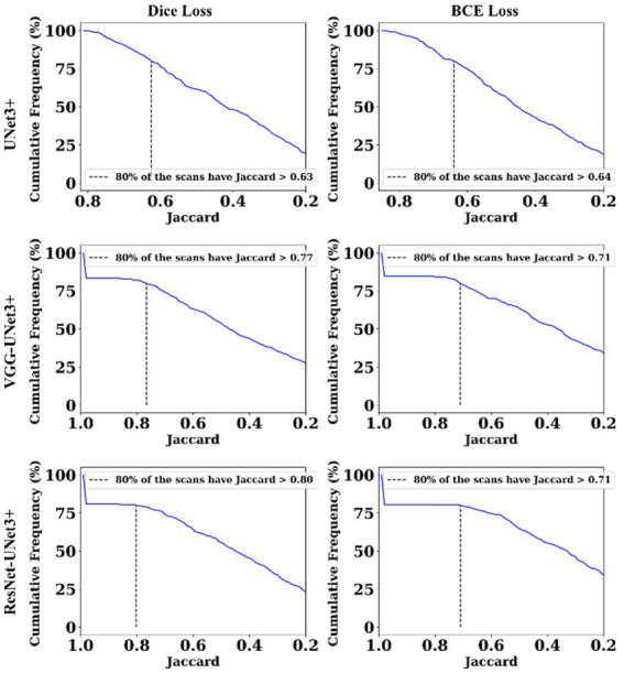
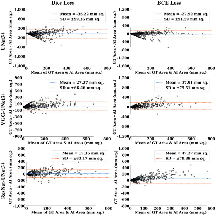
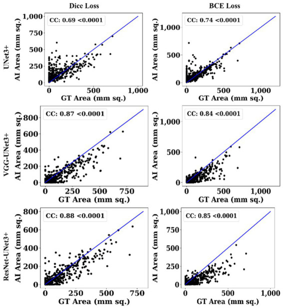
Annotations from expert radiologists were used to train one SDL (UNet3+), and two HDL models, namely, VGG-UNet3+ and ResNet-UNet3+. For accuracy, 5-fold cross-validation protocols, training on 3,500 CT scans, and testing on unseen 500 CT scans were adopted in the cloud framework. Two kinds of loss functions were used: Dice Similarity (DS) and binary cross-entropy (BCE). Performance was evaluated using (i) Area error, (ii) DS, (iii) Jaccard Index, (iii) Bland-Altman, and (iv) Correlation plots.

RESULTS

Among the two HDL models, ResNet-UNet3+ was superior to UNet3+ by 17 and 10% for Dice and BCE loss. The models were further compressed using quantization showing a percentage size reduction of 66.76, 36.64, and 46.23%, respectively, for UNet3+, VGG-UNet3+, and ResNet-UNet3+. Its stability and reliability were proved by statistical tests such as the Mann-Whitney, Paired -Test, Wilcoxon test, and Friedman test all of which had a  < 0.001.

CONCLUSION

Full-scale skip connections of UNet3+ with VGG and ResNet in HDL framework proved the hypothesis showing powerful results improving the detection accuracy of COVID-19.

摘要

背景与创新性

当逆转录聚合酶链反应(RT-PCR)在新型冠状病毒肺炎(COVID-19)的早期诊断及病情严重程度判断方面效果不佳时,需要进行计算机断层扫描(CT)来诊断COVID-19,尤其是对于具有高度磨玻璃影、实变及铺路石样改变的患者。放射科医生发现,在CT图像中手动检测病变极具挑战性且十分繁琐。此前尝试过单独的深度学习(SDL),但其性能处于中低水平。本研究提出了两种基于云的量化深度学习UNet3+混合(HDL)模型,该模型纳入了全尺度跳跃连接以增强和改进检测效果。

方法

使用来自专业放射科医生的标注数据训练一个SDL(UNet3+)以及两个HDL模型,即VGG-UNet3+和ResNet-UNet3+。为了确保准确性,在云框架中采用了5折交叉验证方案,在3500例CT扫描图像上进行训练,并在500例未见过的CT扫描图像上进行测试。使用了两种损失函数:骰子相似度(DS)和二元交叉熵(BCE)。通过(i)面积误差、(ii)DS、(iii)杰卡德指数、(iii)布兰德-奥特曼分析以及(iv)相关性图来评估性能。

结果

在两种HDL模型中,ResNet-UNet3+在骰子损失和BCE损失方面分别比UNet3+高出17%和10%。通过量化进一步压缩模型,结果显示UNet3+、VGG-UNet3+和ResNet-UNet3+模型的大小分别减少了66.76%、36.64%和46.23%。通过诸如曼-惠特尼检验、配对检验、威尔科克森检验和弗里德曼检验等统计测试证明了其稳定性和可靠性,所有这些检验的P值均<0.001。

结论

在HDL框架中,UNet3+与VGG和ResNet的全尺度跳跃连接证明了该假设,显示出强大的效果,提高了COVID-19的检测准确性。

https://cdn.ncbi.nlm.nih.gov/pmc/blobs/9b31/11240867/e3adc2a833ad/frai-07-1304483-g011.jpg
https://cdn.ncbi.nlm.nih.gov/pmc/blobs/9b31/11240867/7239ac1bc4cf/frai-07-1304483-g001.jpg
https://cdn.ncbi.nlm.nih.gov/pmc/blobs/9b31/11240867/1272cc37fa65/frai-07-1304483-g002.jpg
https://cdn.ncbi.nlm.nih.gov/pmc/blobs/9b31/11240867/4782f5ec9daf/frai-07-1304483-g003.jpg
https://cdn.ncbi.nlm.nih.gov/pmc/blobs/9b31/11240867/aa289da9a969/frai-07-1304483-g004.jpg
https://cdn.ncbi.nlm.nih.gov/pmc/blobs/9b31/11240867/ca429631438d/frai-07-1304483-g005.jpg
https://cdn.ncbi.nlm.nih.gov/pmc/blobs/9b31/11240867/6fa25f4567b4/frai-07-1304483-g006.jpg
https://cdn.ncbi.nlm.nih.gov/pmc/blobs/9b31/11240867/fa534b7755fb/frai-07-1304483-g007.jpg
https://cdn.ncbi.nlm.nih.gov/pmc/blobs/9b31/11240867/a39fa0e29b7d/frai-07-1304483-g008.jpg
https://cdn.ncbi.nlm.nih.gov/pmc/blobs/9b31/11240867/06689bea44c6/frai-07-1304483-g009.jpg
https://cdn.ncbi.nlm.nih.gov/pmc/blobs/9b31/11240867/91355b413c21/frai-07-1304483-g010.jpg
https://cdn.ncbi.nlm.nih.gov/pmc/blobs/9b31/11240867/e3adc2a833ad/frai-07-1304483-g011.jpg

相似文献

1
COVLIAS 3.0: cloud-based quantized hybrid UNet3+ deep learning for COVID-19 lesion detection in lung computed tomography.COVLIAS 3.0:基于云的量化混合UNet3+深度学习用于肺部计算机断层扫描中的新冠肺炎病变检测
Front Artif Intell. 2024 Jun 28;7:1304483. doi: 10.3389/frai.2024.1304483. eCollection 2024.
2
Multicenter Study on COVID-19 Lung Computed Tomography Segmentation with varying Glass Ground Opacities using Unseen Deep Learning Artificial Intelligence Paradigms: COVLIAS 1.0 Validation.多中心研究使用未见深度学习人工智能范例的 COVID-19 肺部计算机断层扫描分割,伴有不同玻璃状混浊:COVLIAS 1.0 验证。
J Med Syst. 2022 Aug 21;46(10):62. doi: 10.1007/s10916-022-01850-y.
3
COVLIAS 1.0 vs. MedSeg: An Artificial Intelligence Framework for Automated Lesion Segmentation in COVID-19 Lung Computed Tomography Scans.COVLIAS 1.0与MedSeg:一种用于COVID-19肺部计算机断层扫描中病变自动分割的人工智能框架。
Diagnostics (Basel). 2022 May 21;12(5):1283. doi: 10.3390/diagnostics12051283.
4
COVLIAS 1.0: Lung Segmentation in COVID-19 Computed Tomography Scans Using Hybrid Deep Learning Artificial Intelligence Models.COVLIAS 1.0:使用混合深度学习人工智能模型对新冠肺炎计算机断层扫描进行肺部分割
Diagnostics (Basel). 2021 Aug 4;11(8):1405. doi: 10.3390/diagnostics11081405.
5
COVLIAS 2.0-cXAI: Cloud-Based Explainable Deep Learning System for COVID-19 Lesion Localization in Computed Tomography Scans.COVLIAS 2.0-cXAI:用于计算机断层扫描中新冠病毒病变定位的基于云的可解释深度学习系统。
Diagnostics (Basel). 2022 Jun 16;12(6):1482. doi: 10.3390/diagnostics12061482.
6
COVLIAS 1.0 vs. MedSeg: Artificial Intelligence-Based Comparative Study for Automated COVID-19 Computed Tomography Lung Segmentation in Italian and Croatian Cohorts.COVLIAS 1.0与MedSeg:意大利和克罗地亚队列中基于人工智能的COVID-19计算机断层扫描肺部分割的比较研究
Diagnostics (Basel). 2021 Dec 15;11(12):2367. doi: 10.3390/diagnostics11122367.
7
Eight pruning deep learning models for low storage and high-speed COVID-19 computed tomography lung segmentation and heatmap-based lesion localization: A multicenter study using COVLIAS 2.0.用于低存储和高速 COVID-19 计算机断层扫描肺分割和基于热图的病变定位的八种剪枝深度学习模型:使用 COVLIAS 2.0 的多中心研究。
Comput Biol Med. 2022 Jul;146:105571. doi: 10.1016/j.compbiomed.2022.105571. Epub 2022 May 21.
8
Inter-Variability Study of COVLIAS 1.0: Hybrid Deep Learning Models for COVID-19 Lung Segmentation in Computed Tomography.COVLIAS 1.0的变异性研究:用于计算机断层扫描中COVID-19肺部分割的混合深度学习模型
Diagnostics (Basel). 2021 Nov 1;11(11):2025. doi: 10.3390/diagnostics11112025.
9
Hybrid U-Net-based deep learning model for volume segmentation of lung nodules in CT images.基于 Hybrid U-Net 的深度学习模型,用于 CT 图像中肺结节的体积分割。
Med Phys. 2022 Nov;49(11):7287-7302. doi: 10.1002/mp.15810. Epub 2022 Aug 17.
10
Ensemble Deep Learning Derived from Transfer Learning for Classification of COVID-19 Patients on Hybrid Deep-Learning-Based Lung Segmentation: A Data Augmentation and Balancing Framework.基于迁移学习的集成深度学习用于基于混合深度学习的肺部分割对新冠肺炎患者进行分类:一种数据增强与平衡框架
Diagnostics (Basel). 2023 Jun 2;13(11):1954. doi: 10.3390/diagnostics13111954.

本文引用的文献

1
A Pharmaceutical Paradigm for Cardiovascular Composite Risk Assessment Using Novel Radiogenomics Risk Predictors in Precision Explainable Artificial Intelligence Framework: Clinical Trial Tool.基于新型放射基因组学风险预测因子的精准可解释人工智能框架下心血管复合风险评估的药物治疗范式:临床试验工具。
Front Biosci (Landmark Ed). 2023 Oct 19;28(10):248. doi: 10.31083/j.fbl2810248.
2
Attention-Based UNet Deep Learning Model for Plaque Segmentation in Carotid Ultrasound for Stroke Risk Stratification: An Artificial Intelligence Paradigm.基于注意力机制的U-Net深度学习模型用于颈动脉超声斑块分割以进行中风风险分层:一种人工智能范式
J Cardiovasc Dev Dis. 2022 Sep 27;9(10):326. doi: 10.3390/jcdd9100326.
3
Segmentation-Based Classification Deep Learning Model Embedded with Explainable AI for COVID-19 Detection in Chest X-ray Scans.
基于分割的分类深度学习模型,嵌入可解释人工智能用于胸部X光扫描中的新冠肺炎检测
Diagnostics (Basel). 2022 Sep 2;12(9):2132. doi: 10.3390/diagnostics12092132.
4
Vascular Implications of COVID-19: Role of Radiological Imaging, Artificial Intelligence, and Tissue Characterization: A Special Report.2019冠状病毒病的血管影响:放射影像学、人工智能和组织特征分析的作用:特别报告
J Cardiovasc Dev Dis. 2022 Aug 15;9(8):268. doi: 10.3390/jcdd9080268.
5
Multicenter Study on COVID-19 Lung Computed Tomography Segmentation with varying Glass Ground Opacities using Unseen Deep Learning Artificial Intelligence Paradigms: COVLIAS 1.0 Validation.多中心研究使用未见深度学习人工智能范例的 COVID-19 肺部计算机断层扫描分割,伴有不同玻璃状混浊:COVLIAS 1.0 验证。
J Med Syst. 2022 Aug 21;46(10):62. doi: 10.1007/s10916-022-01850-y.
6
Eight pruning deep learning models for low storage and high-speed COVID-19 computed tomography lung segmentation and heatmap-based lesion localization: A multicenter study using COVLIAS 2.0.用于低存储和高速 COVID-19 计算机断层扫描肺分割和基于热图的病变定位的八种剪枝深度学习模型:使用 COVLIAS 2.0 的多中心研究。
Comput Biol Med. 2022 Jul;146:105571. doi: 10.1016/j.compbiomed.2022.105571. Epub 2022 May 21.
7
COVLIAS 2.0-cXAI: Cloud-Based Explainable Deep Learning System for COVID-19 Lesion Localization in Computed Tomography Scans.COVLIAS 2.0-cXAI:用于计算机断层扫描中新冠病毒病变定位的基于云的可解释深度学习系统。
Diagnostics (Basel). 2022 Jun 16;12(6):1482. doi: 10.3390/diagnostics12061482.
8
Automatic geographic atrophy segmentation using optical attenuation in OCT scans with deep learning.利用深度学习通过光学衰减在光学相干断层扫描(OCT)中自动进行地理萎缩分割
Biomed Opt Express. 2022 Feb 7;13(3):1328-1343. doi: 10.1364/BOE.449314. eCollection 2022 Mar 1.
9
An Efficient Defocus Blur Segmentation Scheme Based on Hybrid LTP and PCNN.基于混合 LTP 和 PCNN 的高效离焦模糊分割方案。
Sensors (Basel). 2022 Apr 1;22(7):2724. doi: 10.3390/s22072724.
10
An artificial intelligence framework and its bias for brain tumor segmentation: A narrative review.一种用于脑肿瘤分割的人工智能框架及其偏差:一篇综述。
Comput Biol Med. 2022 Apr;143:105273. doi: 10.1016/j.compbiomed.2022.105273. Epub 2022 Feb 19.