• 文献检索
  • 文档翻译
  • 深度研究
  • 学术资讯
  • Suppr Zotero 插件Zotero 插件
  • 邀请有礼
  • 套餐&价格
  • 历史记录
应用&插件
Suppr Zotero 插件Zotero 插件浏览器插件Mac 客户端Windows 客户端微信小程序
定价
高级版会员购买积分包购买API积分包
服务
文献检索文档翻译深度研究API 文档MCP 服务
关于我们
关于 Suppr公司介绍联系我们用户协议隐私条款
关注我们

Suppr 超能文献

核心技术专利:CN118964589B侵权必究
粤ICP备2023148730 号-1Suppr @ 2026

文献检索

告别复杂PubMed语法,用中文像聊天一样搜索,搜遍4000万医学文献。AI智能推荐,让科研检索更轻松。

立即免费搜索

文件翻译

保留排版,准确专业,支持PDF/Word/PPT等文件格式,支持 12+语言互译。

免费翻译文档

深度研究

AI帮你快速写综述,25分钟生成高质量综述,智能提取关键信息,辅助科研写作。

立即免费体验

COVLIAS 1.0的变异性研究:用于计算机断层扫描中COVID-19肺部分割的混合深度学习模型

Inter-Variability Study of COVLIAS 1.0: Hybrid Deep Learning Models for COVID-19 Lung Segmentation in Computed Tomography.

作者信息

Suri Jasjit S, Agarwal Sushant, Elavarthi Pranav, Pathak Rajesh, Ketireddy Vedmanvitha, Columbu Marta, Saba Luca, Gupta Suneet K, Faa Gavino, Singh Inder M, Turk Monika, Chadha Paramjit S, Johri Amer M, Khanna Narendra N, Viskovic Klaudija, Mavrogeni Sophie, Laird John R, Pareek Gyan, Miner Martin, Sobel David W, Balestrieri Antonella, Sfikakis Petros P, Tsoulfas George, Protogerou Athanasios, Misra Durga Prasanna, Agarwal Vikas, Kitas George D, Teji Jagjit S, Al-Maini Mustafa, Dhanjil Surinder K, Nicolaides Andrew, Sharma Aditya, Rathore Vijay, Fatemi Mostafa, Alizad Azra, Krishnan Pudukode R, Ferenc Nagy, Ruzsa Zoltan, Gupta Archna, Naidu Subbaram, Kalra Mannudeep K

机构信息

Stroke Diagnostic and Monitoring Division, AtheroPoint™, Roseville, CA 95661, USA.

Advanced Knowledge Engineering Centre, GBTI, Roseville, CA 95661, USA.

出版信息

Diagnostics (Basel). 2021 Nov 1;11(11):2025. doi: 10.3390/diagnostics11112025.

DOI:10.3390/diagnostics11112025
PMID:34829372
原文链接:https://pmc.ncbi.nlm.nih.gov/articles/PMC8625039/
Abstract

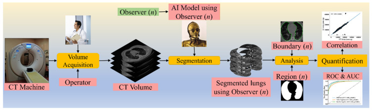
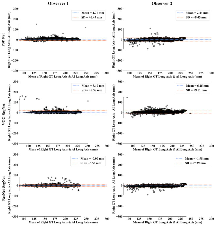
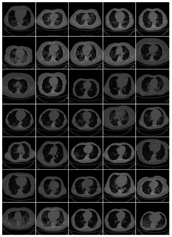
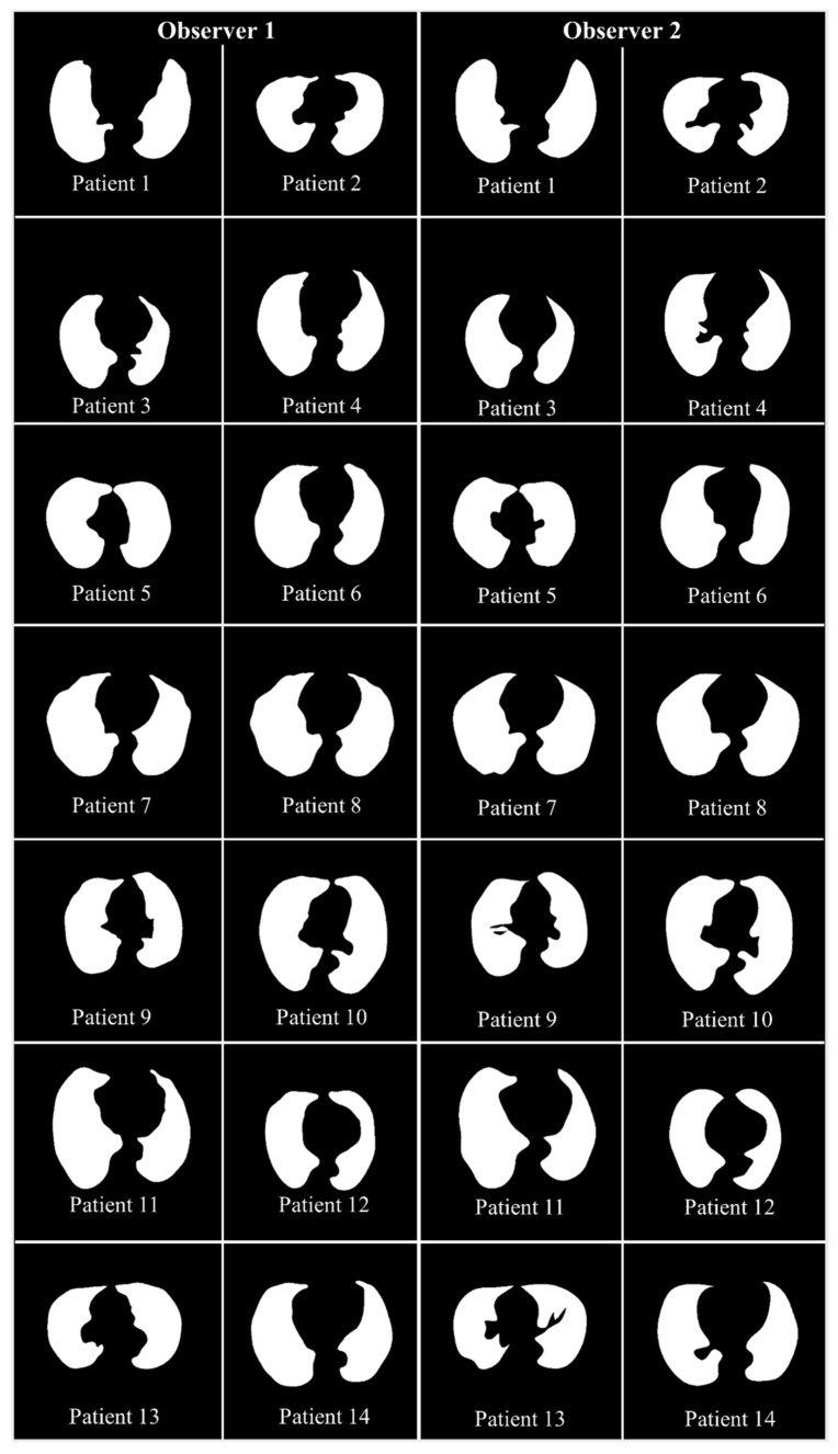
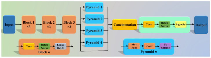
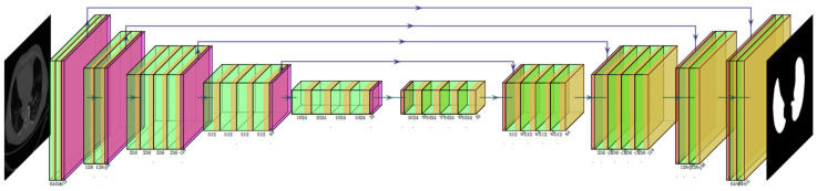
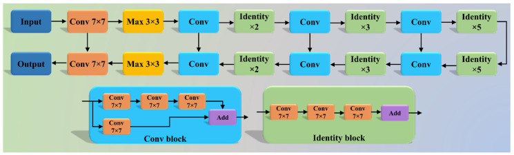
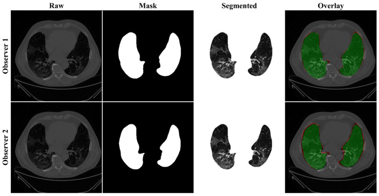
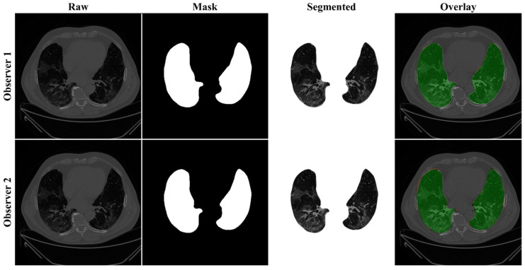
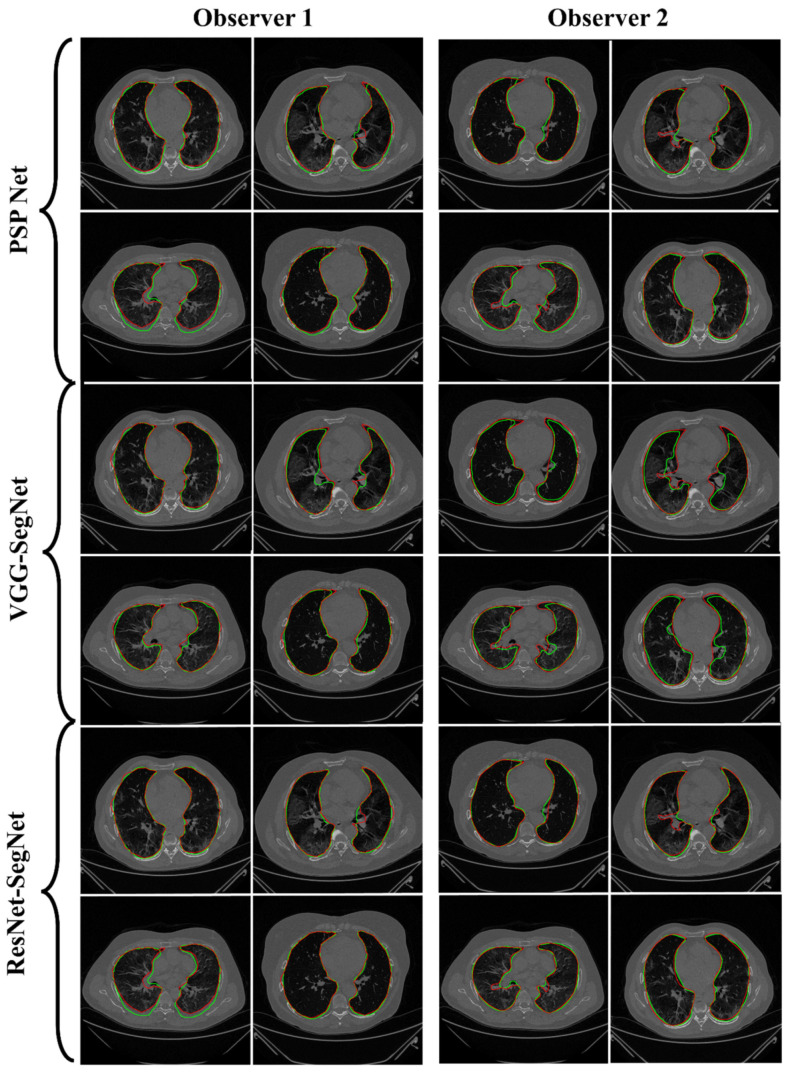
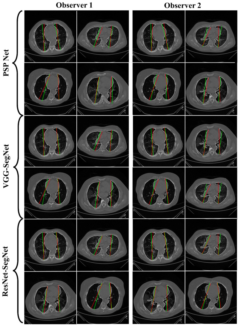
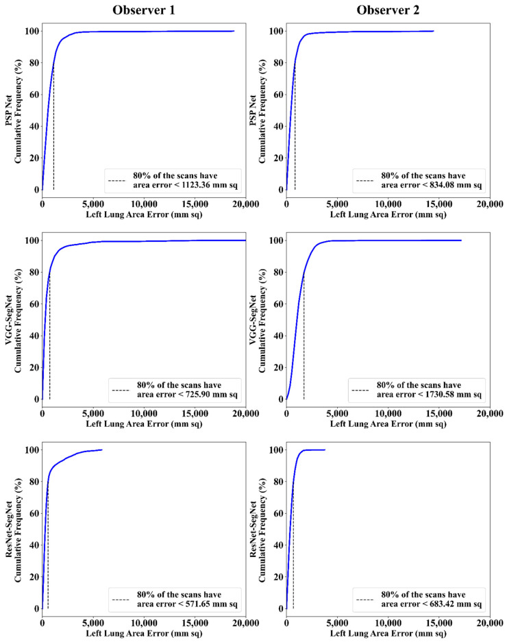
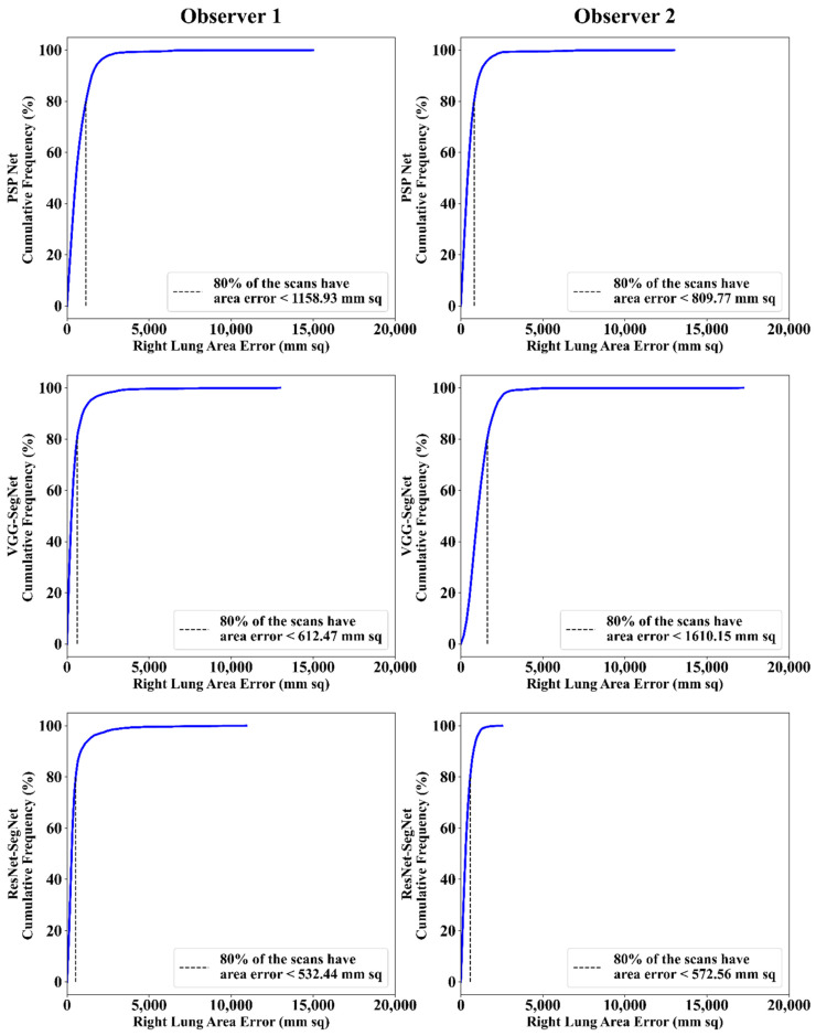
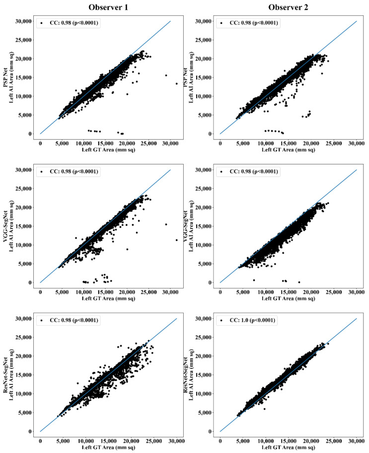
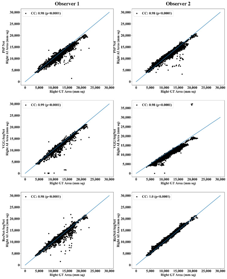
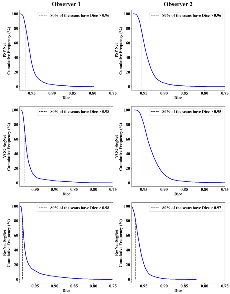
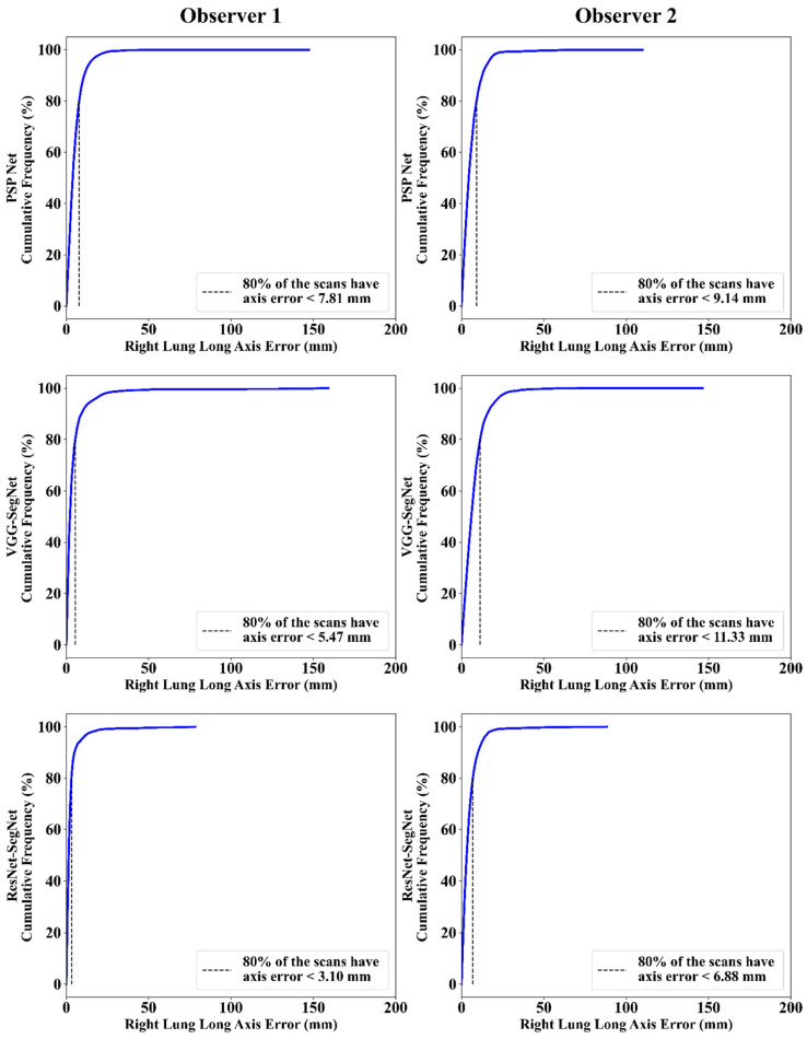
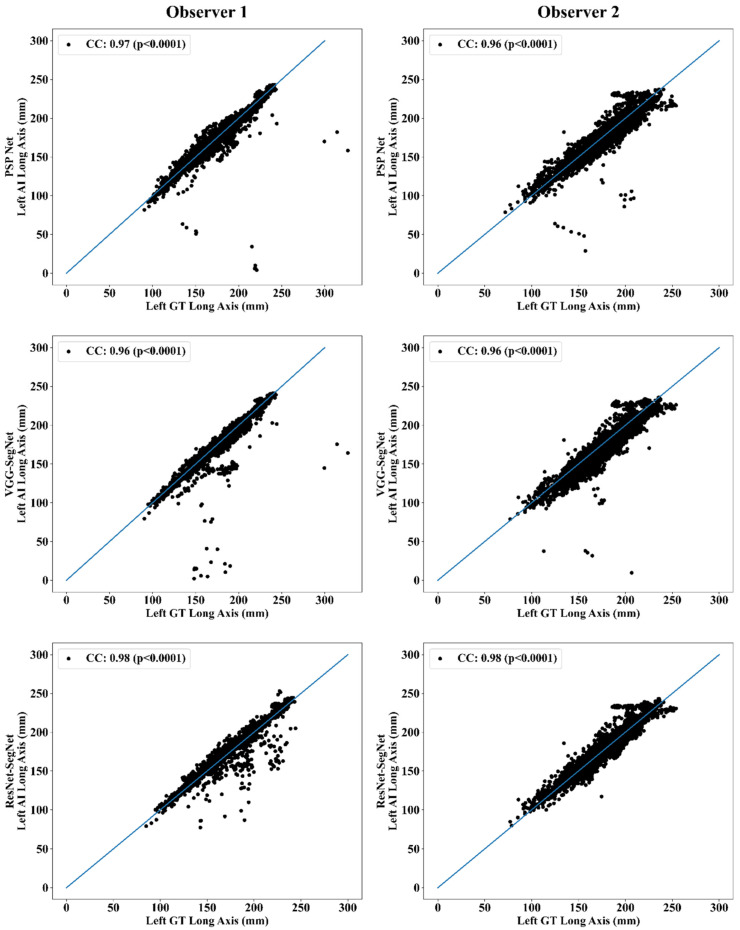
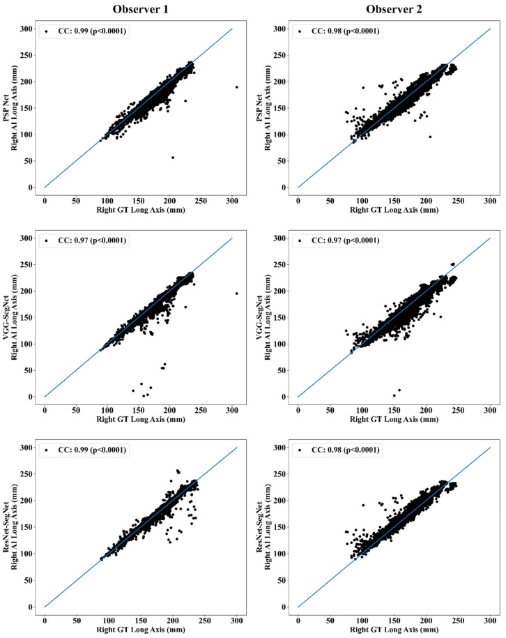
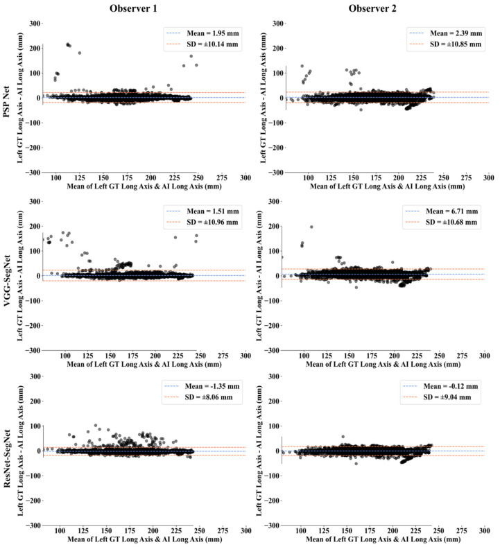
: For COVID-19 lung severity, segmentation of lungs on computed tomography (CT) is the first crucial step. Current deep learning (DL)-based Artificial Intelligence (AI) models have a bias in the training stage of segmentation because only one set of ground truth (GT) annotations are evaluated. We propose a robust and stable inter-variability analysis of CT lung segmentation in COVID-19 to avoid the effect of bias. : The proposed inter-variability study consists of two GT tracers for lung segmentation on chest CT. Three AI models, PSP Net, VGG-SegNet, and ResNet-SegNet, were trained using GT annotations. We hypothesized that if AI models are trained on the GT tracings from multiple experience levels, and if the AI performance on the test data between these AI models is within the 5% range, one can consider such an AI model robust and unbiased. The K5 protocol (training to testing: 80%:20%) was adapted. Ten kinds of metrics were used for performance evaluation. : The database consisted of 5000 CT chest images from 72 COVID-19-infected patients. By computing the coefficient of correlations (CC) between the output of the two AI models trained corresponding to the two GT tracers, computing their differences in their CC, and repeating the process for all three AI-models, we show the differences as 0%, 0.51%, and 2.04% (all < 5%), thereby validating the hypothesis. The performance was comparable; however, it had the following order: ResNet-SegNet > PSP Net > VGG-SegNet. : The AI models were clinically robust and stable during the inter-variability analysis on the CT lung segmentation on COVID-19 patients.

摘要

对于新冠肺炎的肺部严重程度,在计算机断层扫描(CT)上对肺部进行分割是至关重要的第一步。当前基于深度学习(DL)的人工智能(AI)模型在分割训练阶段存在偏差,因为仅评估了一组地面真值(GT)注释。我们提出了一种针对新冠肺炎CT肺部分割的稳健且稳定的变异间分析,以避免偏差的影响。:所提出的变异间研究包括用于胸部CT肺部分割的两个GT追踪器。使用GT注释训练了三个AI模型,即PSP Net、VGG - SegNet和ResNet - SegNet。我们假设,如果AI模型在来自多个经验水平的GT追踪上进行训练,并且如果这些AI模型之间在测试数据上的AI性能在5%范围内,则可以认为这样的AI模型是稳健且无偏差的。采用了K5协议(训练与测试比例:80%:20%)。使用了十种指标进行性能评估。:该数据库由来自72名新冠肺炎感染患者的5000张胸部CT图像组成。通过计算对应于两个GT追踪器训练的两个AI模型输出之间的相关系数(CC),计算它们在CC上的差异,并对所有三个AI模型重复该过程,我们显示差异为0%、0.51%和2.04%(均<5%),从而验证了该假设。性能具有可比性;然而,其顺序如下:ResNet - SegNet > PSP Net > VGG - SegNet。:在对新冠肺炎患者的CT肺部分割进行变异间分析期间,AI模型在临床上是稳健且稳定的。

https://cdn.ncbi.nlm.nih.gov/pmc/blobs/9b3b/8625039/357e3098e0a2/diagnostics-11-02025-g026.jpg
https://cdn.ncbi.nlm.nih.gov/pmc/blobs/9b3b/8625039/626263ba8383/diagnostics-11-02025-g001.jpg
https://cdn.ncbi.nlm.nih.gov/pmc/blobs/9b3b/8625039/da7b425e8129/diagnostics-11-02025-g002.jpg
https://cdn.ncbi.nlm.nih.gov/pmc/blobs/9b3b/8625039/c760f0ab1d0a/diagnostics-11-02025-g003.jpg
https://cdn.ncbi.nlm.nih.gov/pmc/blobs/9b3b/8625039/02bcf8f17304/diagnostics-11-02025-g004.jpg
https://cdn.ncbi.nlm.nih.gov/pmc/blobs/9b3b/8625039/16f1a3cc6e8c/diagnostics-11-02025-g005.jpg
https://cdn.ncbi.nlm.nih.gov/pmc/blobs/9b3b/8625039/a299ecafd237/diagnostics-11-02025-g006.jpg
https://cdn.ncbi.nlm.nih.gov/pmc/blobs/9b3b/8625039/f633f5b09fa4/diagnostics-11-02025-g007.jpg
https://cdn.ncbi.nlm.nih.gov/pmc/blobs/9b3b/8625039/1ed0aca48f58/diagnostics-11-02025-g008.jpg
https://cdn.ncbi.nlm.nih.gov/pmc/blobs/9b3b/8625039/12df20c02365/diagnostics-11-02025-g009.jpg
https://cdn.ncbi.nlm.nih.gov/pmc/blobs/9b3b/8625039/b6340dadf45b/diagnostics-11-02025-g010.jpg
https://cdn.ncbi.nlm.nih.gov/pmc/blobs/9b3b/8625039/e37ee1b97aef/diagnostics-11-02025-g011.jpg
https://cdn.ncbi.nlm.nih.gov/pmc/blobs/9b3b/8625039/42e88af48b9e/diagnostics-11-02025-g012.jpg
https://cdn.ncbi.nlm.nih.gov/pmc/blobs/9b3b/8625039/26c9c644bd5f/diagnostics-11-02025-g013.jpg
https://cdn.ncbi.nlm.nih.gov/pmc/blobs/9b3b/8625039/2939ea5dc63c/diagnostics-11-02025-g014.jpg
https://cdn.ncbi.nlm.nih.gov/pmc/blobs/9b3b/8625039/3739f648c1bd/diagnostics-11-02025-g015.jpg
https://cdn.ncbi.nlm.nih.gov/pmc/blobs/9b3b/8625039/ee72f2af8892/diagnostics-11-02025-g016.jpg
https://cdn.ncbi.nlm.nih.gov/pmc/blobs/9b3b/8625039/eec0911b45b9/diagnostics-11-02025-g017.jpg
https://cdn.ncbi.nlm.nih.gov/pmc/blobs/9b3b/8625039/85f1de3b403a/diagnostics-11-02025-g018.jpg
https://cdn.ncbi.nlm.nih.gov/pmc/blobs/9b3b/8625039/99ddfd6965fc/diagnostics-11-02025-g019.jpg
https://cdn.ncbi.nlm.nih.gov/pmc/blobs/9b3b/8625039/780decc98285/diagnostics-11-02025-g020.jpg
https://cdn.ncbi.nlm.nih.gov/pmc/blobs/9b3b/8625039/2f43ff5a0880/diagnostics-11-02025-g021.jpg
https://cdn.ncbi.nlm.nih.gov/pmc/blobs/9b3b/8625039/5e9c200a1414/diagnostics-11-02025-g022.jpg
https://cdn.ncbi.nlm.nih.gov/pmc/blobs/9b3b/8625039/e36f43b8fd7b/diagnostics-11-02025-g023.jpg
https://cdn.ncbi.nlm.nih.gov/pmc/blobs/9b3b/8625039/6163d2f8eaad/diagnostics-11-02025-g024.jpg
https://cdn.ncbi.nlm.nih.gov/pmc/blobs/9b3b/8625039/9e30ea82dce0/diagnostics-11-02025-g025.jpg
https://cdn.ncbi.nlm.nih.gov/pmc/blobs/9b3b/8625039/357e3098e0a2/diagnostics-11-02025-g026.jpg

相似文献

1
Inter-Variability Study of COVLIAS 1.0: Hybrid Deep Learning Models for COVID-19 Lung Segmentation in Computed Tomography.COVLIAS 1.0的变异性研究:用于计算机断层扫描中COVID-19肺部分割的混合深度学习模型
Diagnostics (Basel). 2021 Nov 1;11(11):2025. doi: 10.3390/diagnostics11112025.
2
COVLIAS 1.0: Lung Segmentation in COVID-19 Computed Tomography Scans Using Hybrid Deep Learning Artificial Intelligence Models.COVLIAS 1.0:使用混合深度学习人工智能模型对新冠肺炎计算机断层扫描进行肺部分割
Diagnostics (Basel). 2021 Aug 4;11(8):1405. doi: 10.3390/diagnostics11081405.
3
Multicenter Study on COVID-19 Lung Computed Tomography Segmentation with varying Glass Ground Opacities using Unseen Deep Learning Artificial Intelligence Paradigms: COVLIAS 1.0 Validation.多中心研究使用未见深度学习人工智能范例的 COVID-19 肺部计算机断层扫描分割,伴有不同玻璃状混浊:COVLIAS 1.0 验证。
J Med Syst. 2022 Aug 21;46(10):62. doi: 10.1007/s10916-022-01850-y.
4
COVLIAS 1.0 vs. MedSeg: Artificial Intelligence-Based Comparative Study for Automated COVID-19 Computed Tomography Lung Segmentation in Italian and Croatian Cohorts.COVLIAS 1.0与MedSeg:意大利和克罗地亚队列中基于人工智能的COVID-19计算机断层扫描肺部分割的比较研究
Diagnostics (Basel). 2021 Dec 15;11(12):2367. doi: 10.3390/diagnostics11122367.
5
COVLIAS 1.0 vs. MedSeg: An Artificial Intelligence Framework for Automated Lesion Segmentation in COVID-19 Lung Computed Tomography Scans.COVLIAS 1.0与MedSeg:一种用于COVID-19肺部计算机断层扫描中病变自动分割的人工智能框架。
Diagnostics (Basel). 2022 May 21;12(5):1283. doi: 10.3390/diagnostics12051283.
6
Eight pruning deep learning models for low storage and high-speed COVID-19 computed tomography lung segmentation and heatmap-based lesion localization: A multicenter study using COVLIAS 2.0.用于低存储和高速 COVID-19 计算机断层扫描肺分割和基于热图的病变定位的八种剪枝深度学习模型:使用 COVLIAS 2.0 的多中心研究。
Comput Biol Med. 2022 Jul;146:105571. doi: 10.1016/j.compbiomed.2022.105571. Epub 2022 May 21.
7
COVLIAS 2.0-cXAI: Cloud-Based Explainable Deep Learning System for COVID-19 Lesion Localization in Computed Tomography Scans.COVLIAS 2.0-cXAI:用于计算机断层扫描中新冠病毒病变定位的基于云的可解释深度学习系统。
Diagnostics (Basel). 2022 Jun 16;12(6):1482. doi: 10.3390/diagnostics12061482.
8
Two-stage deep learning model for fully automated pancreas segmentation on computed tomography: Comparison with intra-reader and inter-reader reliability at full and reduced radiation dose on an external dataset.基于 CT 的全自动胰腺分割的两阶段深度学习模型:在外部数据集上比较全剂量和低剂量下的同读者和异读者可靠性。
Med Phys. 2021 May;48(5):2468-2481. doi: 10.1002/mp.14782. Epub 2021 Mar 16.
9
COVID-19 lung CT image segmentation using deep learning methods: U-Net versus SegNet.使用深度学习方法进行COVID-19肺部CT图像分割:U-Net与SegNet对比
BMC Med Imaging. 2021 Feb 9;21(1):19. doi: 10.1186/s12880-020-00529-5.
10
Hybrid deep learning segmentation models for atherosclerotic plaque in internal carotid artery B-mode ultrasound.用于颈内动脉B型超声中动脉粥样硬化斑块的混合深度学习分割模型
Comput Biol Med. 2021 Sep;136:104721. doi: 10.1016/j.compbiomed.2021.104721. Epub 2021 Aug 2.

引用本文的文献

1
Polygenic Risk Score for Cardiovascular Diseases in Artificial Intelligence Paradigm: A Review.人工智能范式中心血管疾病的多基因风险评分:综述。
J Korean Med Sci. 2023 Nov 27;38(46):e395. doi: 10.3346/jkms.2023.38.e395.
2
Cardiovascular disease/stroke risk stratification in deep learning framework: a review.深度学习框架下的心血管疾病/中风风险分层:综述
Cardiovasc Diagn Ther. 2023 Jun 30;13(3):557-598. doi: 10.21037/cdt-22-438. Epub 2023 Jun 5.
3
Ensemble Deep Learning Derived from Transfer Learning for Classification of COVID-19 Patients on Hybrid Deep-Learning-Based Lung Segmentation: A Data Augmentation and Balancing Framework.

本文引用的文献

1
Artificial intelligence-based hybrid deep learning models for image classification: The first narrative review.基于人工智能的混合深度学习模型在图像分类中的应用:第一篇综述性文章
Comput Biol Med. 2021 Oct;137:104803. doi: 10.1016/j.compbiomed.2021.104803. Epub 2021 Aug 27.
2
COVLIAS 1.0: Lung Segmentation in COVID-19 Computed Tomography Scans Using Hybrid Deep Learning Artificial Intelligence Models.COVLIAS 1.0:使用混合深度学习人工智能模型对新冠肺炎计算机断层扫描进行肺部分割
Diagnostics (Basel). 2021 Aug 4;11(8):1405. doi: 10.3390/diagnostics11081405.
3
Multimodality carotid plaque tissue characterization and classification in the artificial intelligence paradigm: a narrative review for stroke application.
基于迁移学习的集成深度学习用于基于混合深度学习的肺部分割对新冠肺炎患者进行分类:一种数据增强与平衡框架
Diagnostics (Basel). 2023 Jun 2;13(11):1954. doi: 10.3390/diagnostics13111954.
4
Economics of Artificial Intelligence in Healthcare: Diagnosis vs. Treatment.医疗保健领域人工智能的经济学:诊断与治疗
Healthcare (Basel). 2022 Dec 9;10(12):2493. doi: 10.3390/healthcare10122493.
5
Recommender System for the Efficient Treatment of COVID-19 Using a Convolutional Neural Network Model and Image Similarity.基于卷积神经网络模型和图像相似度的新冠高效治疗推荐系统
Diagnostics (Basel). 2022 Nov 5;12(11):2700. doi: 10.3390/diagnostics12112700.
6
Editorial on Special Issue "Artificial Intelligence in Image-Based Screening, Diagnostics, and Clinical Care of Cardiopulmonary Diseases".关于“人工智能在心肺疾病基于图像的筛查、诊断和临床护理中的应用”特刊的社论
Diagnostics (Basel). 2022 Oct 27;12(11):2615. doi: 10.3390/diagnostics12112615.
7
Attention-Based UNet Deep Learning Model for Plaque Segmentation in Carotid Ultrasound for Stroke Risk Stratification: An Artificial Intelligence Paradigm.基于注意力机制的U-Net深度学习模型用于颈动脉超声斑块分割以进行中风风险分层:一种人工智能范式
J Cardiovasc Dev Dis. 2022 Sep 27;9(10):326. doi: 10.3390/jcdd9100326.
8
Brain Tumor Characterization Using Radiogenomics in Artificial Intelligence Framework.在人工智能框架中使用放射基因组学进行脑肿瘤特征描述
Cancers (Basel). 2022 Aug 22;14(16):4052. doi: 10.3390/cancers14164052.
9
Eight pruning deep learning models for low storage and high-speed COVID-19 computed tomography lung segmentation and heatmap-based lesion localization: A multicenter study using COVLIAS 2.0.用于低存储和高速 COVID-19 计算机断层扫描肺分割和基于热图的病变定位的八种剪枝深度学习模型:使用 COVLIAS 2.0 的多中心研究。
Comput Biol Med. 2022 Jul;146:105571. doi: 10.1016/j.compbiomed.2022.105571. Epub 2022 May 21.
10
COVLIAS 2.0-cXAI: Cloud-Based Explainable Deep Learning System for COVID-19 Lesion Localization in Computed Tomography Scans.COVLIAS 2.0-cXAI:用于计算机断层扫描中新冠病毒病变定位的基于云的可解释深度学习系统。
Diagnostics (Basel). 2022 Jun 16;12(6):1482. doi: 10.3390/diagnostics12061482.
人工智能范式下的多模态颈动脉斑块组织特征分析与分类:针对卒中应用的叙述性综述
Ann Transl Med. 2021 Jul;9(14):1206. doi: 10.21037/atm-20-7676.
4
Systematic Review of Artificial Intelligence in Acute Respiratory Distress Syndrome for COVID-19 Lung Patients: A Biomedical Imaging Perspective.COVID-19 肺患者急性呼吸窘迫综合征人工智能的系统评价:生物医学成像视角。
IEEE J Biomed Health Inform. 2021 Nov;25(11):4128-4139. doi: 10.1109/JBHI.2021.3103839. Epub 2021 Nov 5.
5
Hybrid deep learning segmentation models for atherosclerotic plaque in internal carotid artery B-mode ultrasound.用于颈内动脉B型超声中动脉粥样硬化斑块的混合深度学习分割模型
Comput Biol Med. 2021 Sep;136:104721. doi: 10.1016/j.compbiomed.2021.104721. Epub 2021 Aug 2.
6
A Means of Assessing Deep Learning-Based Detection of ICOS Protein Expression in Colon Cancer.一种评估基于深度学习的结肠癌中诱导性共刺激分子(ICOS)蛋白表达检测的方法。
Cancers (Basel). 2021 Jul 29;13(15):3825. doi: 10.3390/cancers13153825.
7
Complications in COVID-19 patients: Characteristics of pulmonary embolism.COVID-19 患者的并发症:肺栓塞的特征。
Clin Imaging. 2021 Sep;77:244-249. doi: 10.1016/j.clinimag.2021.05.016. Epub 2021 May 18.
8
Lung Infection Segmentation for COVID-19 Pneumonia Based on a Cascade Convolutional Network from CT Images.基于 CT 图像的级联卷积网络的 COVID-19 肺炎肺部感染分割。
Biomed Res Int. 2021 Apr 15;2021:5544742. doi: 10.1155/2021/5544742. eCollection 2021.
9
Deep CNN models for predicting COVID-19 in CT and x-ray images.用于在CT和X光图像中预测新冠肺炎的深度卷积神经网络模型。
J Med Imaging (Bellingham). 2021 Jan;8(Suppl 1):014502. doi: 10.1117/1.JMI.8.S1.014502. Epub 2021 Apr 21.
10
Computed tomography findings of COVID-19 pneumonia in Intensive Care Unit-patients.重症监护病房中新冠肺炎患者的计算机断层扫描结果
J Public Health Res. 2021 Apr 19;10(3):2270. doi: 10.4081/jphr.2021.2270.