• 文献检索
  • 文档翻译
  • 深度研究
  • 学术资讯
  • Suppr Zotero 插件Zotero 插件
  • 邀请有礼
  • 套餐&价格
  • 历史记录
应用&插件
Suppr Zotero 插件Zotero 插件浏览器插件Mac 客户端Windows 客户端微信小程序
定价
高级版会员购买积分包购买API积分包
服务
文献检索文档翻译深度研究API 文档MCP 服务
关于我们
关于 Suppr公司介绍联系我们用户协议隐私条款
关注我们

Suppr 超能文献

核心技术专利:CN118964589B侵权必究
粤ICP备2023148730 号-1Suppr @ 2026

文献检索

告别复杂PubMed语法,用中文像聊天一样搜索,搜遍4000万医学文献。AI智能推荐,让科研检索更轻松。

立即免费搜索

文件翻译

保留排版,准确专业,支持PDF/Word/PPT等文件格式,支持 12+语言互译。

免费翻译文档

深度研究

AI帮你快速写综述,25分钟生成高质量综述,智能提取关键信息,辅助科研写作。

立即免费体验

COVLIAS 2.0-cXAI:用于计算机断层扫描中新冠病毒病变定位的基于云的可解释深度学习系统。

COVLIAS 2.0-cXAI: Cloud-Based Explainable Deep Learning System for COVID-19 Lesion Localization in Computed Tomography Scans.

作者信息

Suri Jasjit S, Agarwal Sushant, Chabert Gian Luca, Carriero Alessandro, Paschè Alessio, Danna Pietro S C, Saba Luca, Mehmedović Armin, Faa Gavino, Singh Inder M, Turk Monika, Chadha Paramjit S, Johri Amer M, Khanna Narendra N, Mavrogeni Sophie, Laird John R, Pareek Gyan, Miner Martin, Sobel David W, Balestrieri Antonella, Sfikakis Petros P, Tsoulfas George, Protogerou Athanasios D, Misra Durga Prasanna, Agarwal Vikas, Kitas George D, Teji Jagjit S, Al-Maini Mustafa, Dhanjil Surinder K, Nicolaides Andrew, Sharma Aditya, Rathore Vijay, Fatemi Mostafa, Alizad Azra, Krishnan Pudukode R, Nagy Ferenc, Ruzsa Zoltan, Fouda Mostafa M, Naidu Subbaram, Viskovic Klaudija, Kalra Mannudeep K

机构信息

Stroke Diagnostic and Monitoring Division, AtheroPoint™, Roseville, CA 95661, USA.

Advanced Knowledge Engineering Centre, GBTI, Roseville, CA 95661, USA.

出版信息

Diagnostics (Basel). 2022 Jun 16;12(6):1482. doi: 10.3390/diagnostics12061482.

DOI:10.3390/diagnostics12061482
PMID:35741292
原文链接:https://pmc.ncbi.nlm.nih.gov/articles/PMC9221733/
Abstract

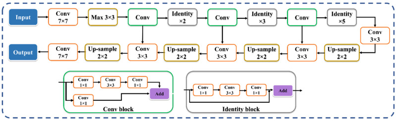
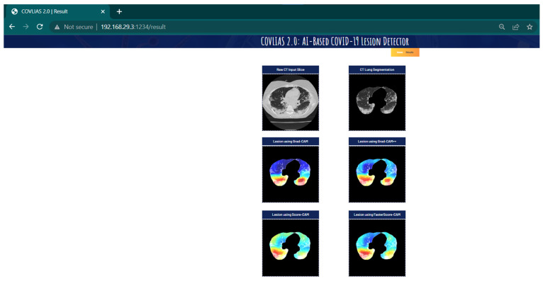
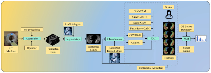
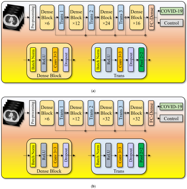
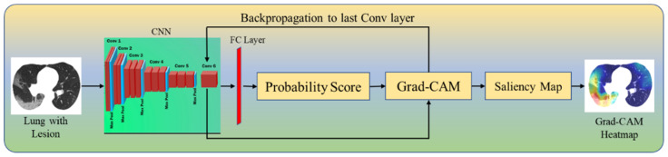
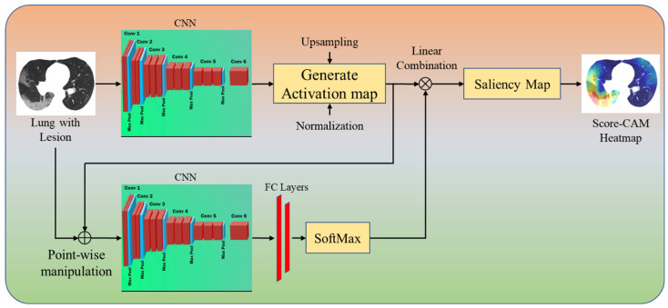
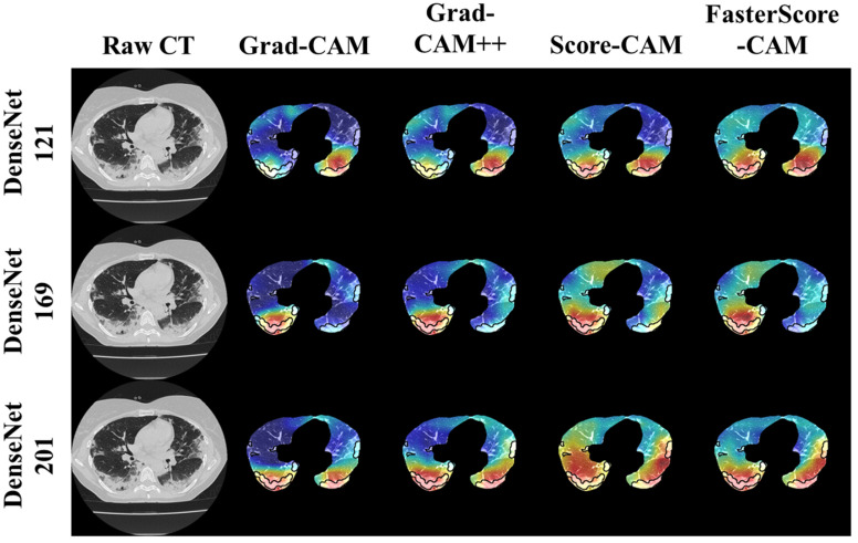
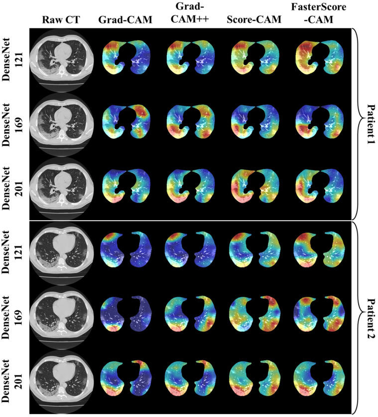
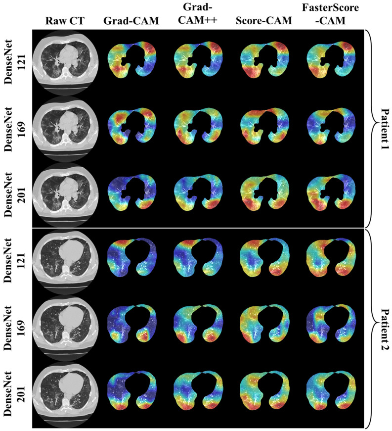
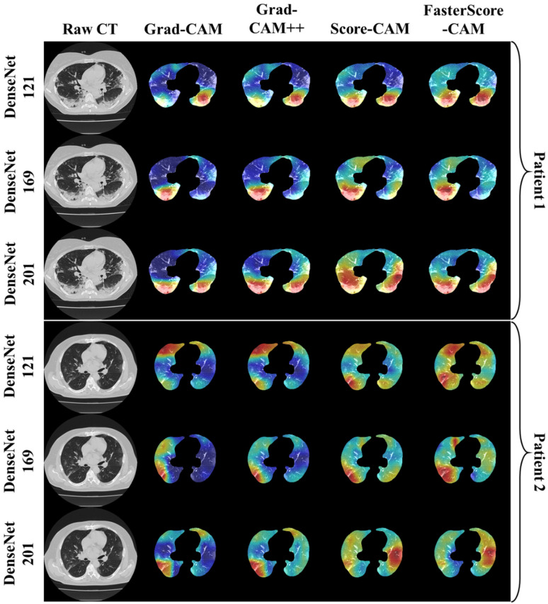
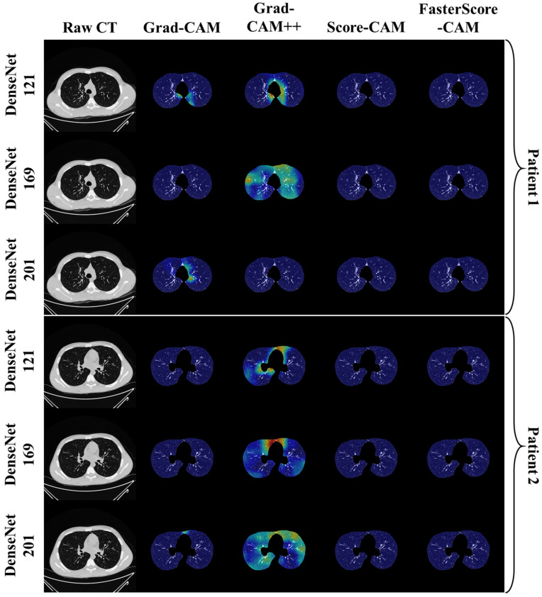
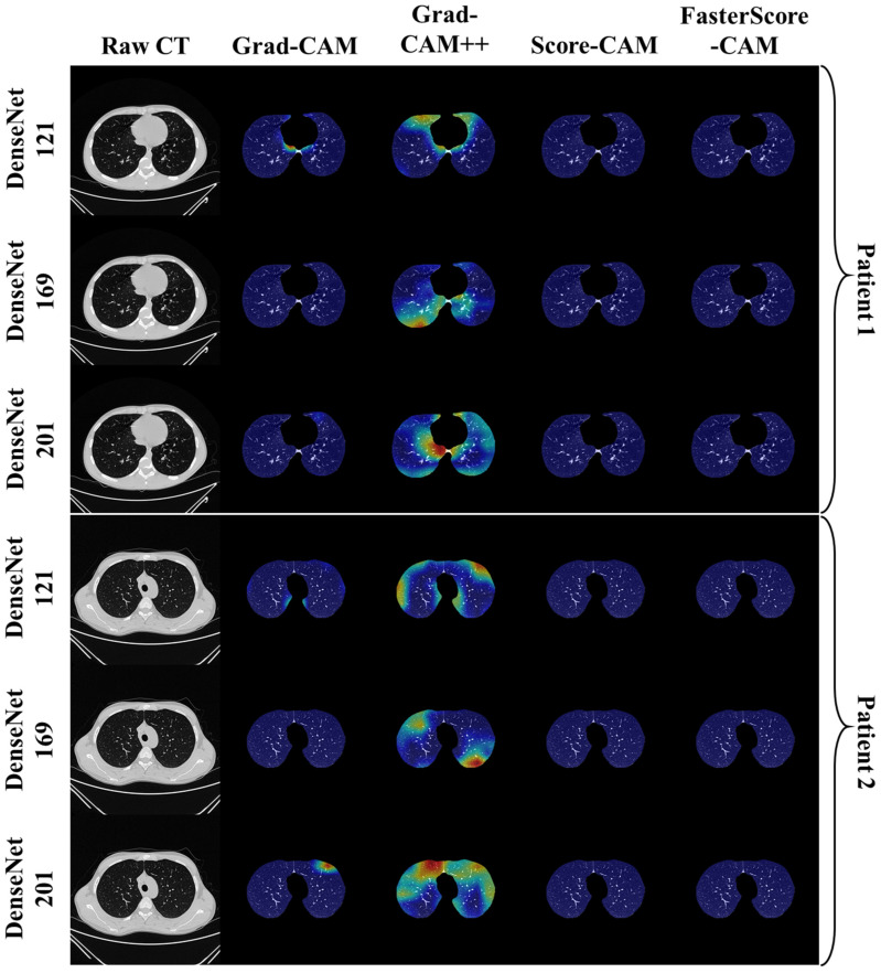
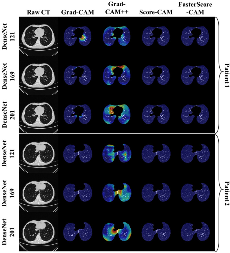
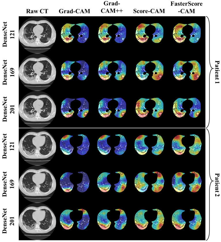
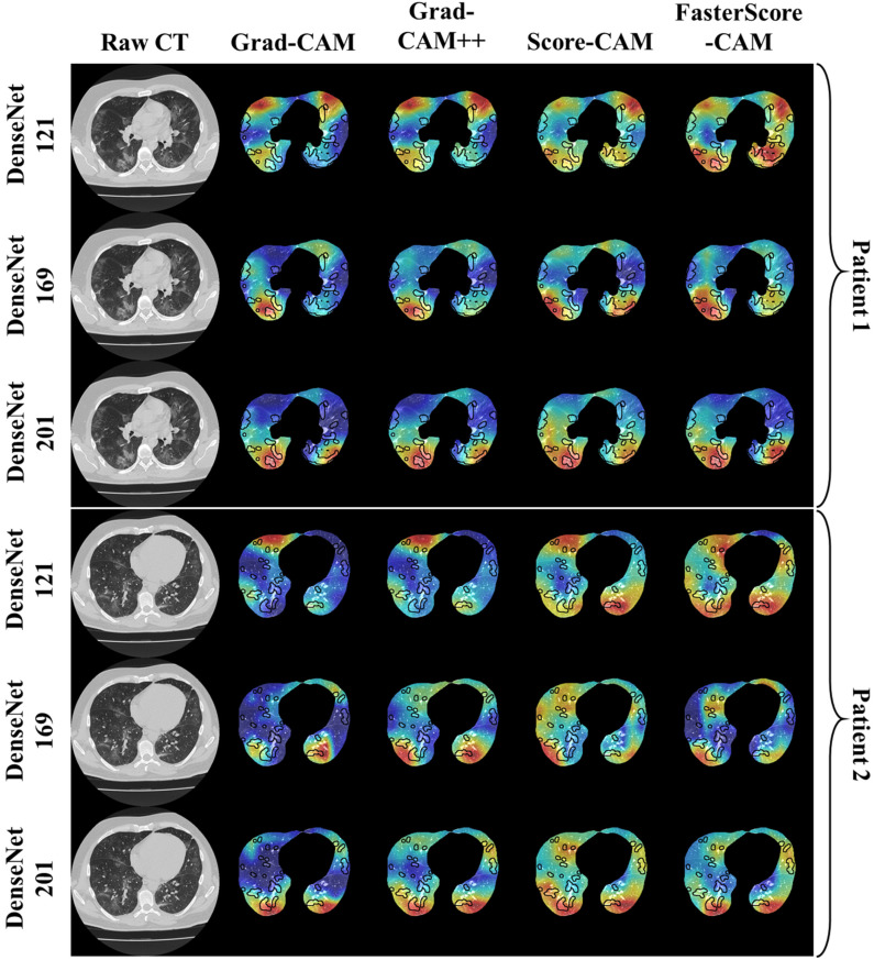
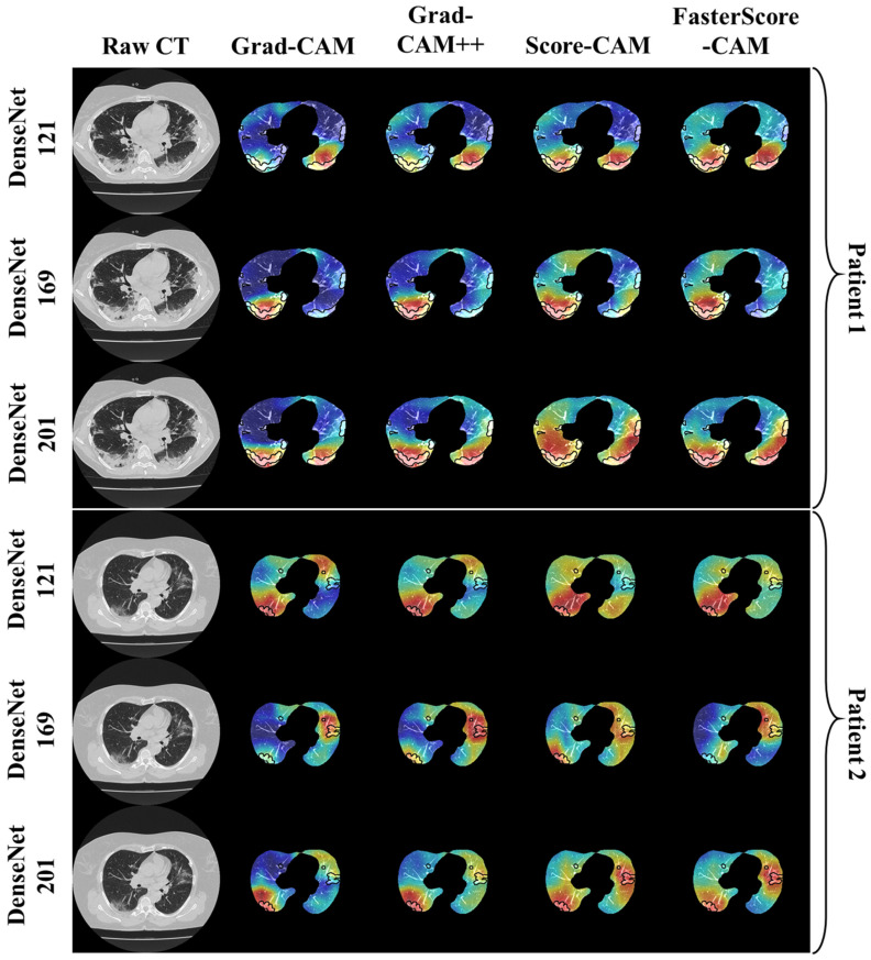
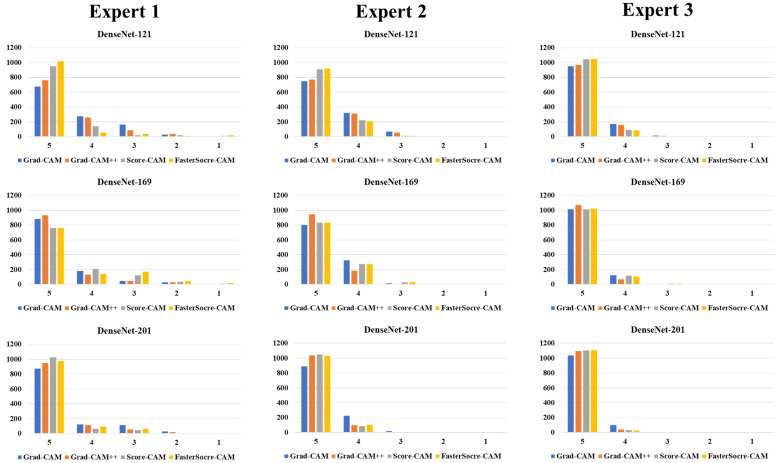
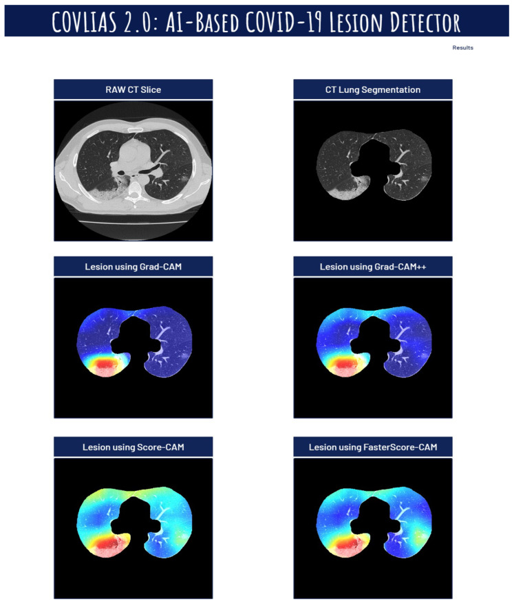
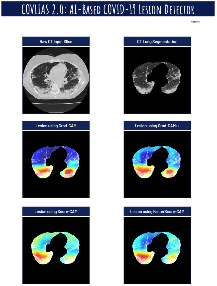
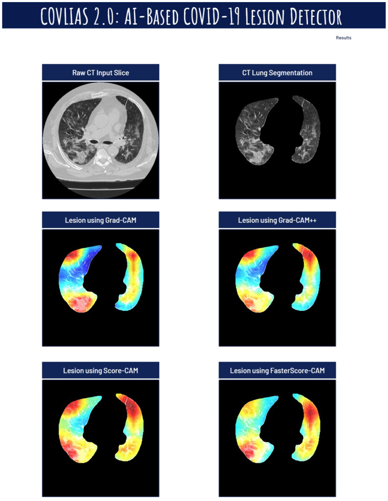
Background: The previous COVID-19 lung diagnosis system lacks both scientific validation and the role of explainable artificial intelligence (AI) for understanding lesion localization. This study presents a cloud-based explainable AI, the “COVLIAS 2.0-cXAI” system using four kinds of class activation maps (CAM) models. Methodology: Our cohort consisted of ~6000 CT slices from two sources (Croatia, 80 COVID-19 patients and Italy, 15 control patients). COVLIAS 2.0-cXAI design consisted of three stages: (i) automated lung segmentation using hybrid deep learning ResNet-UNet model by automatic adjustment of Hounsfield units, hyperparameter optimization, and parallel and distributed training, (ii) classification using three kinds of DenseNet (DN) models (DN-121, DN-169, DN-201), and (iii) validation using four kinds of CAM visualization techniques: gradient-weighted class activation mapping (Grad-CAM), Grad-CAM++, score-weighted CAM (Score-CAM), and FasterScore-CAM. The COVLIAS 2.0-cXAI was validated by three trained senior radiologists for its stability and reliability. The Friedman test was also performed on the scores of the three radiologists. Results: The ResNet-UNet segmentation model resulted in dice similarity of 0.96, Jaccard index of 0.93, a correlation coefficient of 0.99, with a figure-of-merit of 95.99%, while the classifier accuracies for the three DN nets (DN-121, DN-169, and DN-201) were 98%, 98%, and 99% with a loss of ~0.003, ~0.0025, and ~0.002 using 50 epochs, respectively. The mean AUC for all three DN models was 0.99 (p < 0.0001). The COVLIAS 2.0-cXAI showed 80% scans for mean alignment index (MAI) between heatmaps and gold standard, a score of four out of five, establishing the system for clinical settings. Conclusions: The COVLIAS 2.0-cXAI successfully showed a cloud-based explainable AI system for lesion localization in lung CT scans.

摘要

背景

先前的新冠病毒肺炎肺部诊断系统既缺乏科学验证,也缺乏可解释人工智能(AI)在理解病变定位方面的作用。本研究提出了一种基于云的可解释AI,即使用四种类激活映射(CAM)模型的“COVLIAS 2.0-cXAI”系统。方法:我们的队列由来自两个来源的约6000张CT切片组成(克罗地亚,80例新冠病毒肺炎患者;意大利,15例对照患者)。COVLIAS 2.0-cXAI设计包括三个阶段:(i)使用混合深度学习ResNet-UNet模型通过自动调整亨氏单位、超参数优化以及并行和分布式训练进行自动肺部分割,(ii)使用三种DenseNet(DN)模型(DN-121、DN-169、DN-201)进行分类,(iii)使用四种CAM可视化技术进行验证:梯度加权类激活映射(Grad-CAM)、Grad-CAM++、分数加权CAM(Score-CAM)和FasterScore-CAM。COVLIAS 2.0-cXAI由三名训练有素的资深放射科医生对其稳定性和可靠性进行了验证。还对三名放射科医生的评分进行了弗里德曼检验。结果:ResNet-UNet分割模型的骰子相似度为0.96,杰卡德指数为0.93,相关系数为0.99,品质因数为95.99%,而三种DN网络(DN-121、DN-169和DN-201)的分类器准确率分别为98%、98%和99%,在使用50个轮次时损失分别约为0.003、0.0025和0.002。所有三种DN模型的平均AUC为0.99(p<0.0001)。COVLIAS 2.0-cXAI显示,热图与金标准之间的平均对齐指数(MAI)在80%的扫描中得分为五分之四,为临床应用建立了该系统。结论:COVLIAS 2.0-cXAI成功展示了一种用于肺部CT扫描病变定位的基于云的可解释AI系统。

https://cdn.ncbi.nlm.nih.gov/pmc/blobs/c056/9221733/cd483ece3497/diagnostics-12-01482-g022.jpg
https://cdn.ncbi.nlm.nih.gov/pmc/blobs/c056/9221733/cfae8169f260/diagnostics-12-01482-g0A1.jpg
https://cdn.ncbi.nlm.nih.gov/pmc/blobs/c056/9221733/db5d998f4398/diagnostics-12-01482-g001.jpg
https://cdn.ncbi.nlm.nih.gov/pmc/blobs/c056/9221733/00129f2e227a/diagnostics-12-01482-g002.jpg
https://cdn.ncbi.nlm.nih.gov/pmc/blobs/c056/9221733/5c70b8ad850a/diagnostics-12-01482-g003.jpg
https://cdn.ncbi.nlm.nih.gov/pmc/blobs/c056/9221733/e5f861516afa/diagnostics-12-01482-g004a.jpg
https://cdn.ncbi.nlm.nih.gov/pmc/blobs/c056/9221733/0acb64449f4a/diagnostics-12-01482-g005.jpg
https://cdn.ncbi.nlm.nih.gov/pmc/blobs/c056/9221733/15e3f920300d/diagnostics-12-01482-g006.jpg
https://cdn.ncbi.nlm.nih.gov/pmc/blobs/c056/9221733/12ef3323469c/diagnostics-12-01482-g007.jpg
https://cdn.ncbi.nlm.nih.gov/pmc/blobs/c056/9221733/3478f2e7c417/diagnostics-12-01482-g008.jpg
https://cdn.ncbi.nlm.nih.gov/pmc/blobs/c056/9221733/3d6f9477103d/diagnostics-12-01482-g009.jpg
https://cdn.ncbi.nlm.nih.gov/pmc/blobs/c056/9221733/94acd2696e24/diagnostics-12-01482-g010.jpg
https://cdn.ncbi.nlm.nih.gov/pmc/blobs/c056/9221733/27a80e17f1e6/diagnostics-12-01482-g011.jpg
https://cdn.ncbi.nlm.nih.gov/pmc/blobs/c056/9221733/18095696ddbe/diagnostics-12-01482-g012.jpg
https://cdn.ncbi.nlm.nih.gov/pmc/blobs/c056/9221733/0458be332ab2/diagnostics-12-01482-g013.jpg
https://cdn.ncbi.nlm.nih.gov/pmc/blobs/c056/9221733/ad5018a546e3/diagnostics-12-01482-g014.jpg
https://cdn.ncbi.nlm.nih.gov/pmc/blobs/c056/9221733/c65437d2b84b/diagnostics-12-01482-g015.jpg
https://cdn.ncbi.nlm.nih.gov/pmc/blobs/c056/9221733/70c7a1fa03da/diagnostics-12-01482-g016.jpg
https://cdn.ncbi.nlm.nih.gov/pmc/blobs/c056/9221733/f79c7785a981/diagnostics-12-01482-g017.jpg
https://cdn.ncbi.nlm.nih.gov/pmc/blobs/c056/9221733/a41abe21a77f/diagnostics-12-01482-g018.jpg
https://cdn.ncbi.nlm.nih.gov/pmc/blobs/c056/9221733/ec5ec4c63c61/diagnostics-12-01482-g019.jpg
https://cdn.ncbi.nlm.nih.gov/pmc/blobs/c056/9221733/daeb464680a1/diagnostics-12-01482-g020.jpg
https://cdn.ncbi.nlm.nih.gov/pmc/blobs/c056/9221733/1eff53fb1ab1/diagnostics-12-01482-g021.jpg
https://cdn.ncbi.nlm.nih.gov/pmc/blobs/c056/9221733/cd483ece3497/diagnostics-12-01482-g022.jpg

相似文献

1
COVLIAS 2.0-cXAI: Cloud-Based Explainable Deep Learning System for COVID-19 Lesion Localization in Computed Tomography Scans.COVLIAS 2.0-cXAI:用于计算机断层扫描中新冠病毒病变定位的基于云的可解释深度学习系统。
Diagnostics (Basel). 2022 Jun 16;12(6):1482. doi: 10.3390/diagnostics12061482.
2
Multicenter Study on COVID-19 Lung Computed Tomography Segmentation with varying Glass Ground Opacities using Unseen Deep Learning Artificial Intelligence Paradigms: COVLIAS 1.0 Validation.多中心研究使用未见深度学习人工智能范例的 COVID-19 肺部计算机断层扫描分割,伴有不同玻璃状混浊:COVLIAS 1.0 验证。
J Med Syst. 2022 Aug 21;46(10):62. doi: 10.1007/s10916-022-01850-y.
3
Eight pruning deep learning models for low storage and high-speed COVID-19 computed tomography lung segmentation and heatmap-based lesion localization: A multicenter study using COVLIAS 2.0.用于低存储和高速 COVID-19 计算机断层扫描肺分割和基于热图的病变定位的八种剪枝深度学习模型:使用 COVLIAS 2.0 的多中心研究。
Comput Biol Med. 2022 Jul;146:105571. doi: 10.1016/j.compbiomed.2022.105571. Epub 2022 May 21.
4
COVLIAS 1.0: Lung Segmentation in COVID-19 Computed Tomography Scans Using Hybrid Deep Learning Artificial Intelligence Models.COVLIAS 1.0:使用混合深度学习人工智能模型对新冠肺炎计算机断层扫描进行肺部分割
Diagnostics (Basel). 2021 Aug 4;11(8):1405. doi: 10.3390/diagnostics11081405.
5
COVLIAS 1.0 vs. MedSeg: Artificial Intelligence-Based Comparative Study for Automated COVID-19 Computed Tomography Lung Segmentation in Italian and Croatian Cohorts.COVLIAS 1.0与MedSeg:意大利和克罗地亚队列中基于人工智能的COVID-19计算机断层扫描肺部分割的比较研究
Diagnostics (Basel). 2021 Dec 15;11(12):2367. doi: 10.3390/diagnostics11122367.
6
COVLIAS 1.0 vs. MedSeg: An Artificial Intelligence Framework for Automated Lesion Segmentation in COVID-19 Lung Computed Tomography Scans.COVLIAS 1.0与MedSeg:一种用于COVID-19肺部计算机断层扫描中病变自动分割的人工智能框架。
Diagnostics (Basel). 2022 May 21;12(5):1283. doi: 10.3390/diagnostics12051283.
7
COVLIAS 3.0: cloud-based quantized hybrid UNet3+ deep learning for COVID-19 lesion detection in lung computed tomography.COVLIAS 3.0:基于云的量化混合UNet3+深度学习用于肺部计算机断层扫描中的新冠肺炎病变检测
Front Artif Intell. 2024 Jun 28;7:1304483. doi: 10.3389/frai.2024.1304483. eCollection 2024.
8
Segmentation-Based Classification Deep Learning Model Embedded with Explainable AI for COVID-19 Detection in Chest X-ray Scans.基于分割的分类深度学习模型,嵌入可解释人工智能用于胸部X光扫描中的新冠肺炎检测
Diagnostics (Basel). 2022 Sep 2;12(9):2132. doi: 10.3390/diagnostics12092132.
9
Grad-CAM-Based Explainable Artificial Intelligence Related to Medical Text Processing.基于Grad-CAM的与医学文本处理相关的可解释人工智能
Bioengineering (Basel). 2023 Sep 10;10(9):1070. doi: 10.3390/bioengineering10091070.
10
A novel approach of brain-computer interfacing (BCI) and Grad-CAM based explainable artificial intelligence: Use case scenario for smart healthcare.一种新的脑机接口 (BCI) 和基于 Grad-CAM 的可解释人工智能方法:智能医疗保健用例场景。
J Neurosci Methods. 2024 Aug;408:110159. doi: 10.1016/j.jneumeth.2024.110159. Epub 2024 May 7.

引用本文的文献

1
Transformer and Attention-Based Architectures for Segmentation of Coronary Arterial Walls in Intravascular Ultrasound: A Narrative Review.基于Transformer和注意力机制的血管内超声冠状动脉壁分割架构:综述
Diagnostics (Basel). 2025 Mar 26;15(7):848. doi: 10.3390/diagnostics15070848.
2
Explainable AI for Bipolar Disorder Diagnosis Using Hjorth Parameters.使用 Hjorth 参数的双相情感障碍诊断的可解释人工智能。
Diagnostics (Basel). 2025 Jan 29;15(3):316. doi: 10.3390/diagnostics15030316.
3
Deep learning methods for improving the accuracy and efficiency of pathological image analysis.

本文引用的文献

1
Dense Convolutional Network and Its Application in Medical Image Analysis.密集卷积网络及其在医学图像分析中的应用。
Biomed Res Int. 2022 Apr 25;2022:2384830. doi: 10.1155/2022/2384830. eCollection 2022.
2
Explainable AI for CNN-based prostate tumor segmentation in multi-parametric MRI correlated to whole mount histopathology.基于卷积神经网络的多参数 MRI 前列腺肿瘤分割的可解释人工智能与全组织病理相关。
Radiat Oncol. 2022 Apr 2;17(1):65. doi: 10.1186/s13014-022-02035-0.
3
A Powerful Paradigm for Cardiovascular Risk Stratification Using Multiclass, Multi-Label, and Ensemble-Based Machine Learning Paradigms: A Narrative Review.
用于提高病理图像分析准确性和效率的深度学习方法。
Sci Prog. 2025 Jan-Mar;108(1):368504241306830. doi: 10.1177/00368504241306830.
4
An Artificial Intelligence-Based Non-Invasive Approach for Cardiovascular Disease Risk Stratification in Obstructive Sleep Apnea Patients: A Narrative Review.一种基于人工智能的阻塞性睡眠呼吸暂停患者心血管疾病风险分层的非侵入性方法:叙述性综述。
Rev Cardiovasc Med. 2024 Dec 28;25(12):463. doi: 10.31083/j.rcm2512463. eCollection 2024 Dec.
5
UltraAIGenomics: Artificial Intelligence-Based Cardiovascular Disease Risk Assessment by Fusion of Ultrasound-Based Radiomics and Genomics Features for Preventive, Personalized and Precision Medicine: A Narrative Review.超人工智能基因组学:基于超声影像组学和基因组学特征融合的人工智能心血管疾病风险评估用于预防、个性化和精准医学:一篇综述
Rev Cardiovasc Med. 2024 May 22;25(5):184. doi: 10.31083/j.rcm2505184. eCollection 2024 May.
6
Towards Improved XAI-Based Epidemiological Research into the Next Potential Pandemic.迈向基于可解释人工智能的流行病学研究,以应对下一次潜在的大流行。
Life (Basel). 2024 Jun 21;14(7):783. doi: 10.3390/life14070783.
7
Four Transformer-Based Deep Learning Classifiers Embedded with an Attention U-Net-Based Lung Segmenter and Layer-Wise Relevance Propagation-Based Heatmaps for COVID-19 X-ray Scans.四种基于Transformer的深度学习分类器,嵌入了基于注意力U-Net的肺部分割器和基于逐层相关性传播的热图,用于COVID-19 X光扫描。
Diagnostics (Basel). 2024 Jul 16;14(14):1534. doi: 10.3390/diagnostics14141534.
8
COVLIAS 3.0: cloud-based quantized hybrid UNet3+ deep learning for COVID-19 lesion detection in lung computed tomography.COVLIAS 3.0:基于云的量化混合UNet3+深度学习用于肺部计算机断层扫描中的新冠肺炎病变检测
Front Artif Intell. 2024 Jun 28;7:1304483. doi: 10.3389/frai.2024.1304483. eCollection 2024.
9
Comparing Visual and Software-Based Quantitative Assessment Scores of Lungs' Parenchymal Involvement Quantification in COVID-19 Patients.比较COVID-19患者肺部实质受累量化的视觉和基于软件的定量评估分数
Diagnostics (Basel). 2024 May 8;14(10):985. doi: 10.3390/diagnostics14100985.
10
Deep learning approach for cardiovascular disease risk stratification and survival analysis on a Canadian cohort.基于加拿大队列的心血管疾病风险分层和生存分析的深度学习方法
Int J Cardiovasc Imaging. 2024 Jun;40(6):1283-1303. doi: 10.1007/s10554-024-03100-3. Epub 2024 Apr 28.
一种使用多类、多标签和基于集成的机器学习范式进行心血管风险分层的强大范式:叙述性综述。
Diagnostics (Basel). 2022 Mar 16;12(3):722. doi: 10.3390/diagnostics12030722.
4
Four Types of Multiclass Frameworks for Pneumonia Classification and Its Validation in X-ray Scans Using Seven Types of Deep Learning Artificial Intelligence Models.用于肺炎分类的四种多类框架及其在使用七种深度学习人工智能模型的X射线扫描中的验证
Diagnostics (Basel). 2022 Mar 7;12(3):652. doi: 10.3390/diagnostics12030652.
5
Medical Professional Enhancement Using Explainable Artificial Intelligence in Fetal Cardiac Ultrasound Screening.在胎儿心脏超声筛查中使用可解释人工智能提升医学专业水平。
Biomedicines. 2022 Feb 25;10(3):551. doi: 10.3390/biomedicines10030551.
6
COVID-19 Risk Stratification and Mortality Prediction in Hospitalized Indian Patients: Harnessing clinical data for public health benefits.COVID-19 风险分层和住院印度患者的死亡率预测:利用临床数据造福公众健康。
PLoS One. 2022 Mar 17;17(3):e0264785. doi: 10.1371/journal.pone.0264785. eCollection 2022.
7
Role of imaging in rare COVID-19 vaccine multiorgan complications.影像学在罕见的新冠疫苗多器官并发症中的作用
Insights Imaging. 2022 Mar 14;13(1):44. doi: 10.1186/s13244-022-01176-w.
8
Applications of Explainable Artificial Intelligence in Diagnosis and Surgery.可解释人工智能在诊断与手术中的应用。
Diagnostics (Basel). 2022 Jan 19;12(2):237. doi: 10.3390/diagnostics12020237.
9
Non-invasive coronary imaging in patients with COVID-19: A narrative review.COVID-19 患者的非侵入性冠状动脉成像:叙述性综述。
Eur J Radiol. 2022 Apr;149:110188. doi: 10.1016/j.ejrad.2022.110188. Epub 2022 Feb 1.
10
Long-COVID diagnosis: From diagnostic to advanced AI-driven models.长新冠诊断:从诊断到先进的人工智能驱动模型。
Eur J Radiol. 2022 Mar;148:110164. doi: 10.1016/j.ejrad.2022.110164. Epub 2022 Jan 19.