• 文献检索
  • 文档翻译
  • 深度研究
  • 学术资讯
  • Suppr Zotero 插件Zotero 插件
  • 邀请有礼
  • 套餐&价格
  • 历史记录
应用&插件
Suppr Zotero 插件Zotero 插件浏览器插件Mac 客户端Windows 客户端微信小程序
定价
高级版会员购买积分包购买API积分包
服务
文献检索文档翻译深度研究API 文档MCP 服务
关于我们
关于 Suppr公司介绍联系我们用户协议隐私条款
关注我们

Suppr 超能文献

核心技术专利:CN118964589B侵权必究
粤ICP备2023148730 号-1Suppr @ 2026

文献检索

告别复杂PubMed语法,用中文像聊天一样搜索,搜遍4000万医学文献。AI智能推荐,让科研检索更轻松。

立即免费搜索

文件翻译

保留排版,准确专业,支持PDF/Word/PPT等文件格式,支持 12+语言互译。

免费翻译文档

深度研究

AI帮你快速写综述,25分钟生成高质量综述,智能提取关键信息,辅助科研写作。

立即免费体验

COVLIAS 1.0与MedSeg:一种用于COVID-19肺部计算机断层扫描中病变自动分割的人工智能框架。

COVLIAS 1.0 vs. MedSeg: An Artificial Intelligence Framework for Automated Lesion Segmentation in COVID-19 Lung Computed Tomography Scans.

作者信息

Suri Jasjit S, Agarwal Sushant, Chabert Gian Luca, Carriero Alessandro, Paschè Alessio, Danna Pietro S C, Saba Luca, Mehmedović Armin, Faa Gavino, Singh Inder M, Turk Monika, Chadha Paramjit S, Johri Amer M, Khanna Narendra N, Mavrogeni Sophie, Laird John R, Pareek Gyan, Miner Martin, Sobel David W, Balestrieri Antonella, Sfikakis Petros P, Tsoulfas George, Protogerou Athanasios D, Misra Durga Prasanna, Agarwal Vikas, Kitas George D, Teji Jagjit S, Al-Maini Mustafa, Dhanjil Surinder K, Nicolaides Andrew, Sharma Aditya, Rathore Vijay, Fatemi Mostafa, Alizad Azra, Krishnan Pudukode R, Nagy Ferenc, Ruzsa Zoltan, Fouda Mostafa M, Naidu Subbaram, Viskovic Klaudija, Kalra Manudeep K

机构信息

Stroke Diagnostic and Monitoring Division, AtheroPoint™, Roseville, CA 95661, USA.

Advanced Knowledge Engineering Centre, GBTI, Roseville, CA 95661, USA.

出版信息

Diagnostics (Basel). 2022 May 21;12(5):1283. doi: 10.3390/diagnostics12051283.

DOI:10.3390/diagnostics12051283
PMID:35626438
原文链接:https://pmc.ncbi.nlm.nih.gov/articles/PMC9141749/
Abstract

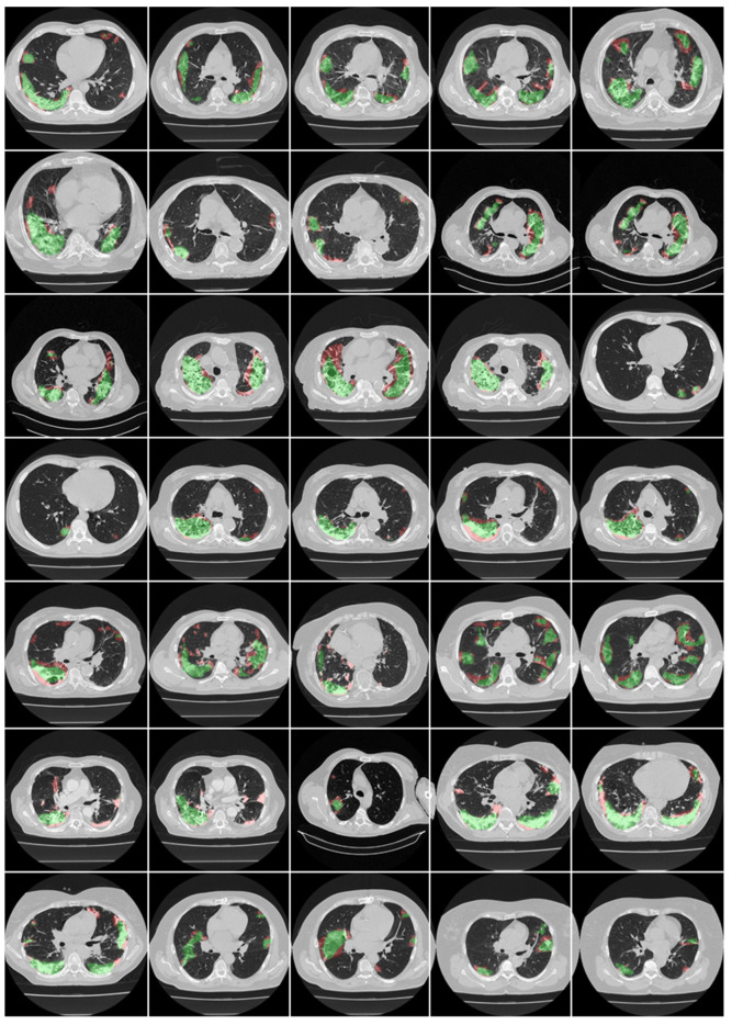
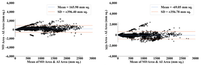
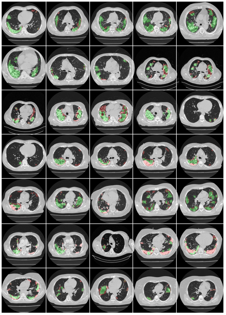
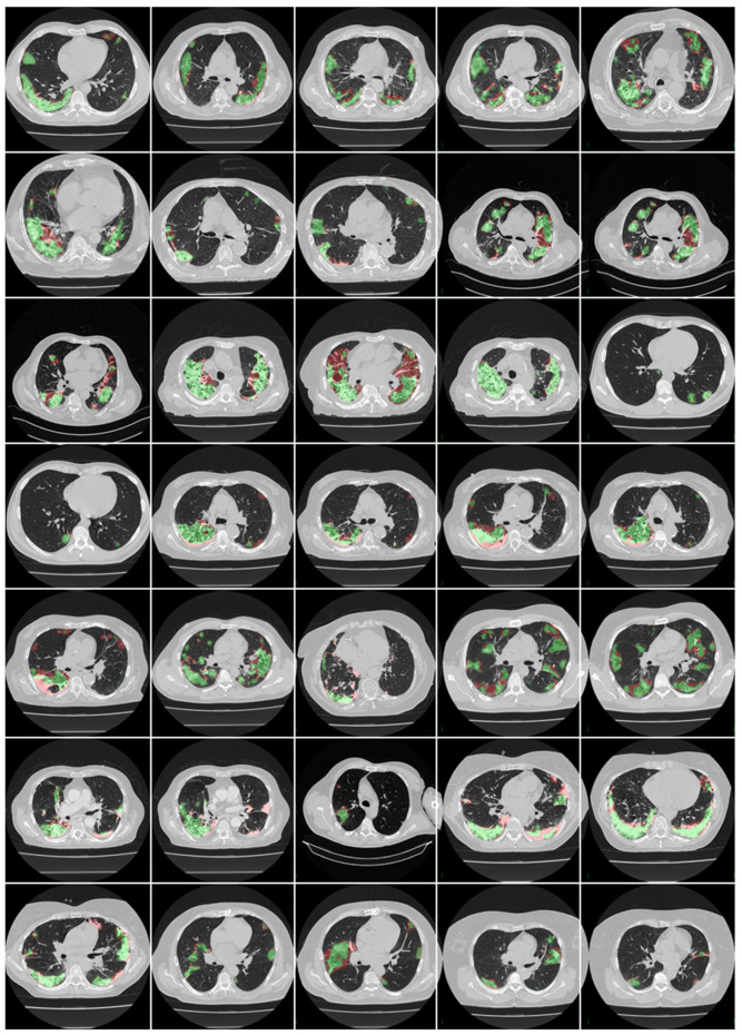
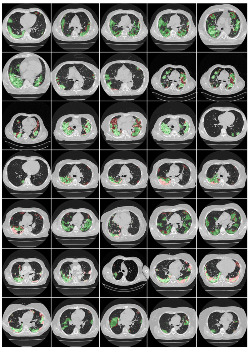
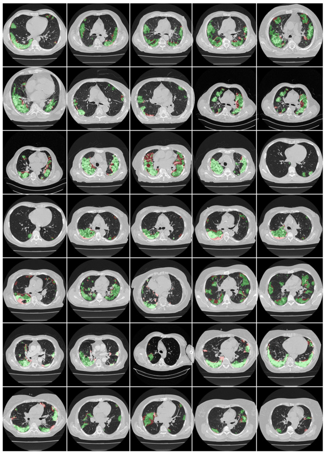
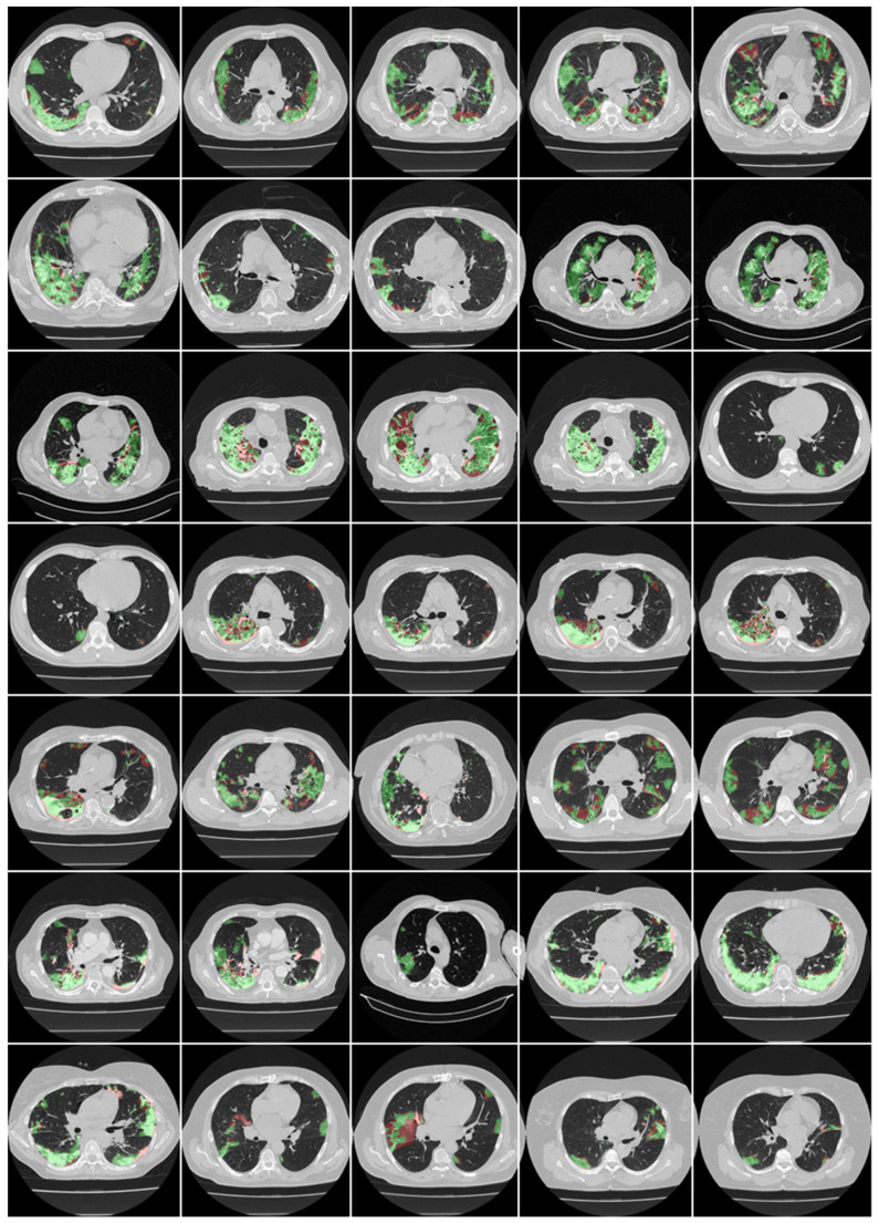
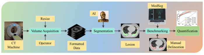
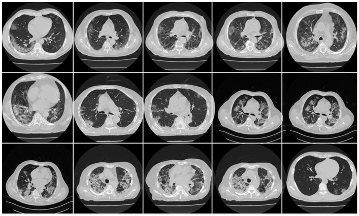
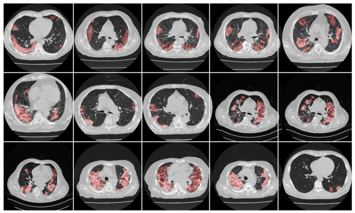
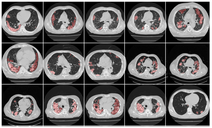
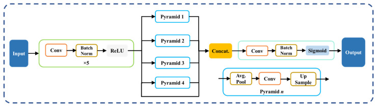
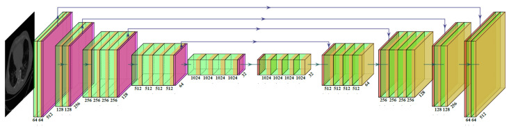
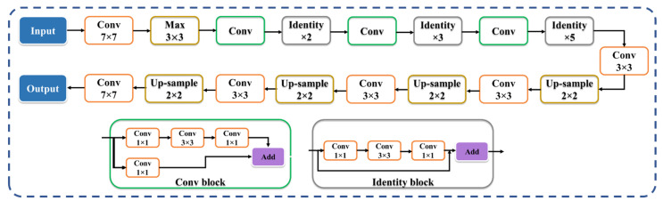
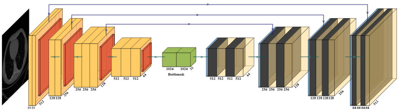
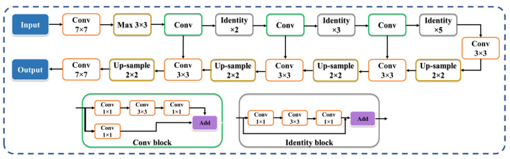
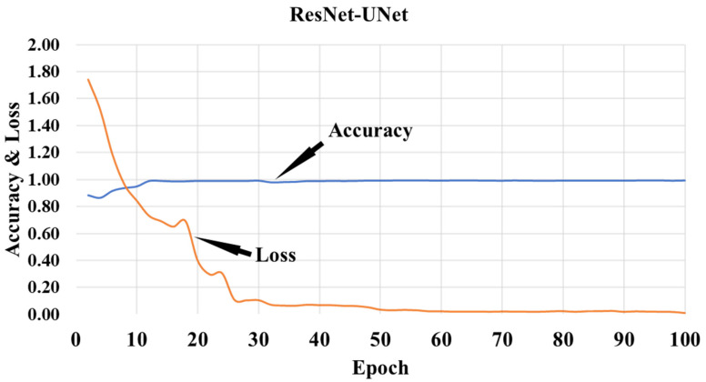
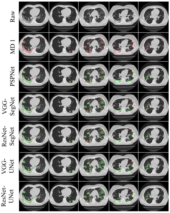
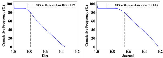
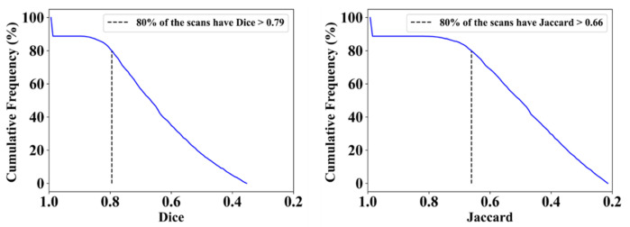
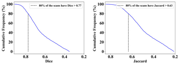
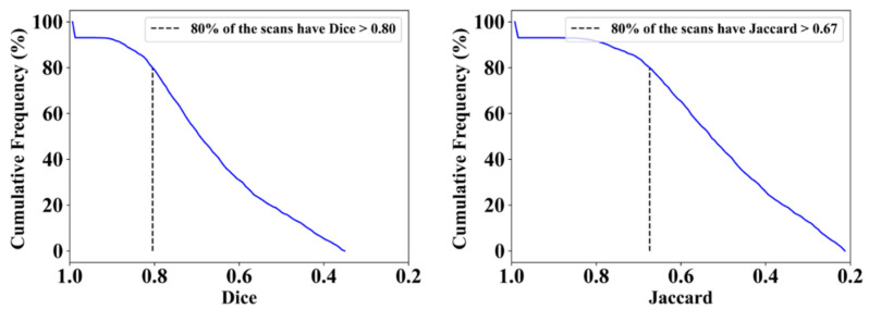
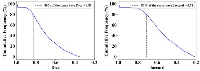
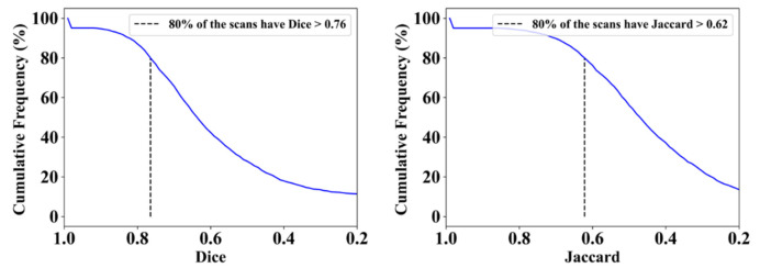
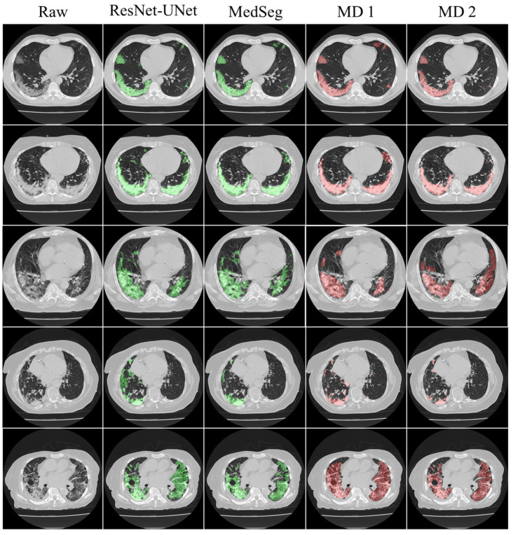
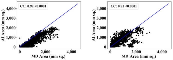
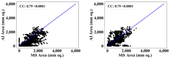
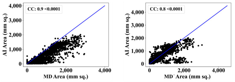
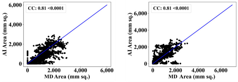
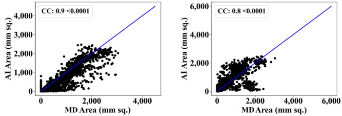
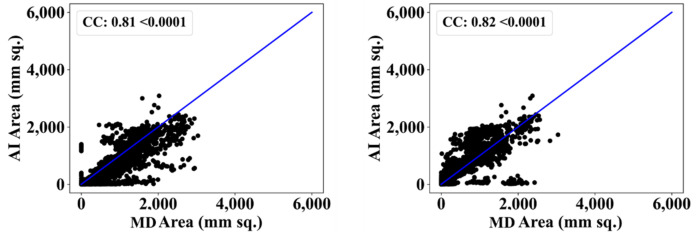
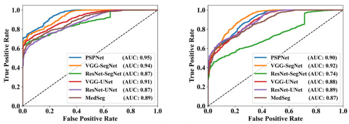
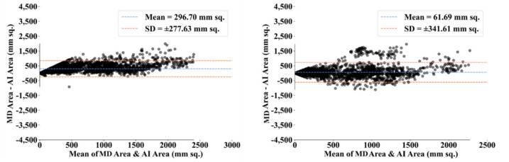
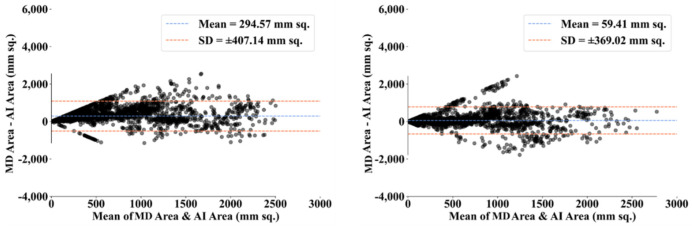
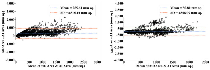
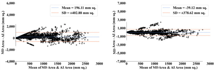
Background: COVID-19 is a disease with multiple variants, and is quickly spreading throughout the world. It is crucial to identify patients who are suspected of having COVID-19 early, because the vaccine is not readily available in certain parts of the world. Methodology: Lung computed tomography (CT) imaging can be used to diagnose COVID-19 as an alternative to the RT-PCR test in some cases. The occurrence of ground-glass opacities in the lung region is a characteristic of COVID-19 in chest CT scans, and these are daunting to locate and segment manually. The proposed study consists of a combination of solo deep learning (DL) and hybrid DL (HDL) models to tackle the lesion location and segmentation more quickly. One DL and four HDL models—namely, PSPNet, VGG-SegNet, ResNet-SegNet, VGG-UNet, and ResNet-UNet—were trained by an expert radiologist. The training scheme adopted a fivefold cross-validation strategy on a cohort of 3000 images selected from a set of 40 COVID-19-positive individuals. Results: The proposed variability study uses tracings from two trained radiologists as part of the validation. Five artificial intelligence (AI) models were benchmarked against MedSeg. The best AI model, ResNet-UNet, was superior to MedSeg by 9% and 15% for Dice and Jaccard, respectively, when compared against MD 1, and by 4% and 8%, respectively, when compared against MD 2. Statistical tests—namely, the Mann−Whitney test, paired t-test, and Wilcoxon test—demonstrated its stability and reliability, with p < 0.0001. The online system for each slice was <1 s. Conclusions: The AI models reliably located and segmented COVID-19 lesions in CT scans. The COVLIAS 1.0Lesion lesion locator passed the intervariability test.

摘要

背景

新型冠状病毒肺炎(COVID-19)是一种具有多种变体的疾病,正在全球迅速传播。尽早识别疑似患有COVID-19的患者至关重要,因为在世界某些地区疫苗尚未广泛可得。方法:在某些情况下,肺部计算机断层扫描(CT)成像可作为逆转录聚合酶链反应(RT-PCR)检测的替代方法用于诊断COVID-19。肺部区域磨玻璃影的出现是胸部CT扫描中COVID-19的一个特征,而手动定位和分割这些磨玻璃影具有挑战性。本研究提出将单深度学习(DL)模型和混合深度学习(HDL)模型相结合,以更快地解决病变定位和分割问题。一位放射科专家训练了一个DL模型和四个HDL模型,即PSPNet、VGG-SegNet、ResNet-SegNet、VGG-UNet和ResNet-UNet。训练方案对从40名COVID-19阳性个体的一组图像中选出的3000幅图像采用五折交叉验证策略。结果:所提出的变异性研究使用了两名训练有素的放射科医生的描记图作为验证的一部分。将五个人工智能(AI)模型与MedSeg进行了基准测试。最佳AI模型ResNet-UNet与MD 1相比,在Dice和Jaccard指标上分别比MedSeg高出9%和15%,与MD 2相比,分别高出4%和8%。统计检验,即曼-惠特尼检验、配对t检验和威尔科克森检验,证明了其稳定性和可靠性,p<0.0001。每幅切片的在线系统用时<1秒。结论:AI模型在CT扫描中可靠地定位和分割了COVID-19病变。COVLIAS 1.0病变定位器通过了变异性测试。

https://cdn.ncbi.nlm.nih.gov/pmc/blobs/ac4f/9141749/66f75920fb89/diagnostics-12-01283-g031.jpg
https://cdn.ncbi.nlm.nih.gov/pmc/blobs/ac4f/9141749/628e10757d56/diagnostics-12-01283-g0A1.jpg
https://cdn.ncbi.nlm.nih.gov/pmc/blobs/ac4f/9141749/c1e670032b9d/diagnostics-12-01283-g0A2.jpg
https://cdn.ncbi.nlm.nih.gov/pmc/blobs/ac4f/9141749/7f3a14d813e4/diagnostics-12-01283-g0A3.jpg
https://cdn.ncbi.nlm.nih.gov/pmc/blobs/ac4f/9141749/ea5a83fbfc80/diagnostics-12-01283-g0A4.jpg
https://cdn.ncbi.nlm.nih.gov/pmc/blobs/ac4f/9141749/829c71c68a3c/diagnostics-12-01283-g0A5.jpg
https://cdn.ncbi.nlm.nih.gov/pmc/blobs/ac4f/9141749/4d80c34b01b3/diagnostics-12-01283-g0A6.jpg
https://cdn.ncbi.nlm.nih.gov/pmc/blobs/ac4f/9141749/23e3dc4afd09/diagnostics-12-01283-g001.jpg
https://cdn.ncbi.nlm.nih.gov/pmc/blobs/ac4f/9141749/9550af0fca2a/diagnostics-12-01283-g002.jpg
https://cdn.ncbi.nlm.nih.gov/pmc/blobs/ac4f/9141749/a64a95c14ba5/diagnostics-12-01283-g003.jpg
https://cdn.ncbi.nlm.nih.gov/pmc/blobs/ac4f/9141749/aea2e9621432/diagnostics-12-01283-g004.jpg
https://cdn.ncbi.nlm.nih.gov/pmc/blobs/ac4f/9141749/a2d239a9fc05/diagnostics-12-01283-g005.jpg
https://cdn.ncbi.nlm.nih.gov/pmc/blobs/ac4f/9141749/5eff587931e6/diagnostics-12-01283-g006.jpg
https://cdn.ncbi.nlm.nih.gov/pmc/blobs/ac4f/9141749/77ad11608660/diagnostics-12-01283-g007.jpg
https://cdn.ncbi.nlm.nih.gov/pmc/blobs/ac4f/9141749/8d20e70305e5/diagnostics-12-01283-g008.jpg
https://cdn.ncbi.nlm.nih.gov/pmc/blobs/ac4f/9141749/c75f7e28aea8/diagnostics-12-01283-g009.jpg
https://cdn.ncbi.nlm.nih.gov/pmc/blobs/ac4f/9141749/fb6183f9c667/diagnostics-12-01283-g010.jpg
https://cdn.ncbi.nlm.nih.gov/pmc/blobs/ac4f/9141749/a80807b19b04/diagnostics-12-01283-g011.jpg
https://cdn.ncbi.nlm.nih.gov/pmc/blobs/ac4f/9141749/25327f86a9a8/diagnostics-12-01283-g012.jpg
https://cdn.ncbi.nlm.nih.gov/pmc/blobs/ac4f/9141749/f9feaa4d8d78/diagnostics-12-01283-g013.jpg
https://cdn.ncbi.nlm.nih.gov/pmc/blobs/ac4f/9141749/bf03aebf5f3b/diagnostics-12-01283-g014.jpg
https://cdn.ncbi.nlm.nih.gov/pmc/blobs/ac4f/9141749/afc84e97cb36/diagnostics-12-01283-g015.jpg
https://cdn.ncbi.nlm.nih.gov/pmc/blobs/ac4f/9141749/66386739b4b0/diagnostics-12-01283-g016.jpg
https://cdn.ncbi.nlm.nih.gov/pmc/blobs/ac4f/9141749/3ae60bf9f606/diagnostics-12-01283-g017.jpg
https://cdn.ncbi.nlm.nih.gov/pmc/blobs/ac4f/9141749/6f193e5ee4ce/diagnostics-12-01283-g018.jpg
https://cdn.ncbi.nlm.nih.gov/pmc/blobs/ac4f/9141749/08fce63e5fdd/diagnostics-12-01283-g019.jpg
https://cdn.ncbi.nlm.nih.gov/pmc/blobs/ac4f/9141749/fcd8e1980699/diagnostics-12-01283-g020.jpg
https://cdn.ncbi.nlm.nih.gov/pmc/blobs/ac4f/9141749/8b883d9fe055/diagnostics-12-01283-g021.jpg
https://cdn.ncbi.nlm.nih.gov/pmc/blobs/ac4f/9141749/d973f4463ca8/diagnostics-12-01283-g022.jpg
https://cdn.ncbi.nlm.nih.gov/pmc/blobs/ac4f/9141749/f954fdaa1691/diagnostics-12-01283-g023.jpg
https://cdn.ncbi.nlm.nih.gov/pmc/blobs/ac4f/9141749/3a30e0ee9bb2/diagnostics-12-01283-g024.jpg
https://cdn.ncbi.nlm.nih.gov/pmc/blobs/ac4f/9141749/1c9d6a3dcbd9/diagnostics-12-01283-g025.jpg
https://cdn.ncbi.nlm.nih.gov/pmc/blobs/ac4f/9141749/21a54b5d1232/diagnostics-12-01283-g026.jpg
https://cdn.ncbi.nlm.nih.gov/pmc/blobs/ac4f/9141749/39ef45bc477e/diagnostics-12-01283-g027.jpg
https://cdn.ncbi.nlm.nih.gov/pmc/blobs/ac4f/9141749/fdbecc18c3e9/diagnostics-12-01283-g028.jpg
https://cdn.ncbi.nlm.nih.gov/pmc/blobs/ac4f/9141749/c8d79c36caca/diagnostics-12-01283-g029.jpg
https://cdn.ncbi.nlm.nih.gov/pmc/blobs/ac4f/9141749/f58a20402e68/diagnostics-12-01283-g030.jpg
https://cdn.ncbi.nlm.nih.gov/pmc/blobs/ac4f/9141749/66f75920fb89/diagnostics-12-01283-g031.jpg

相似文献

1
COVLIAS 1.0 vs. MedSeg: An Artificial Intelligence Framework for Automated Lesion Segmentation in COVID-19 Lung Computed Tomography Scans.COVLIAS 1.0与MedSeg:一种用于COVID-19肺部计算机断层扫描中病变自动分割的人工智能框架。
Diagnostics (Basel). 2022 May 21;12(5):1283. doi: 10.3390/diagnostics12051283.
2
COVLIAS 1.0 vs. MedSeg: Artificial Intelligence-Based Comparative Study for Automated COVID-19 Computed Tomography Lung Segmentation in Italian and Croatian Cohorts.COVLIAS 1.0与MedSeg:意大利和克罗地亚队列中基于人工智能的COVID-19计算机断层扫描肺部分割的比较研究
Diagnostics (Basel). 2021 Dec 15;11(12):2367. doi: 10.3390/diagnostics11122367.
3
Multicenter Study on COVID-19 Lung Computed Tomography Segmentation with varying Glass Ground Opacities using Unseen Deep Learning Artificial Intelligence Paradigms: COVLIAS 1.0 Validation.多中心研究使用未见深度学习人工智能范例的 COVID-19 肺部计算机断层扫描分割,伴有不同玻璃状混浊:COVLIAS 1.0 验证。
J Med Syst. 2022 Aug 21;46(10):62. doi: 10.1007/s10916-022-01850-y.
4
COVLIAS 1.0: Lung Segmentation in COVID-19 Computed Tomography Scans Using Hybrid Deep Learning Artificial Intelligence Models.COVLIAS 1.0:使用混合深度学习人工智能模型对新冠肺炎计算机断层扫描进行肺部分割
Diagnostics (Basel). 2021 Aug 4;11(8):1405. doi: 10.3390/diagnostics11081405.
5
COVLIAS 3.0: cloud-based quantized hybrid UNet3+ deep learning for COVID-19 lesion detection in lung computed tomography.COVLIAS 3.0:基于云的量化混合UNet3+深度学习用于肺部计算机断层扫描中的新冠肺炎病变检测
Front Artif Intell. 2024 Jun 28;7:1304483. doi: 10.3389/frai.2024.1304483. eCollection 2024.
6
Eight pruning deep learning models for low storage and high-speed COVID-19 computed tomography lung segmentation and heatmap-based lesion localization: A multicenter study using COVLIAS 2.0.用于低存储和高速 COVID-19 计算机断层扫描肺分割和基于热图的病变定位的八种剪枝深度学习模型:使用 COVLIAS 2.0 的多中心研究。
Comput Biol Med. 2022 Jul;146:105571. doi: 10.1016/j.compbiomed.2022.105571. Epub 2022 May 21.
7
COVLIAS 2.0-cXAI: Cloud-Based Explainable Deep Learning System for COVID-19 Lesion Localization in Computed Tomography Scans.COVLIAS 2.0-cXAI:用于计算机断层扫描中新冠病毒病变定位的基于云的可解释深度学习系统。
Diagnostics (Basel). 2022 Jun 16;12(6):1482. doi: 10.3390/diagnostics12061482.
8
Inter-Variability Study of COVLIAS 1.0: Hybrid Deep Learning Models for COVID-19 Lung Segmentation in Computed Tomography.COVLIAS 1.0的变异性研究:用于计算机断层扫描中COVID-19肺部分割的混合深度学习模型
Diagnostics (Basel). 2021 Nov 1;11(11):2025. doi: 10.3390/diagnostics11112025.
9
Human-in-the-Loop-A Deep Learning Strategy in Combination with a Patient-Specific Gaussian Mixture Model Leads to the Fast Characterization of Volumetric Ground-Glass Opacity and Consolidation in the Computed Tomography Scans of COVID-19 Patients.人在回路中——一种结合患者特异性高斯混合模型的深度学习策略可快速表征COVID-19患者计算机断层扫描中的磨玻璃密度影和实变。
J Clin Med. 2024 Sep 4;13(17):5231. doi: 10.3390/jcm13175231.
10
Hybrid deep learning segmentation models for atherosclerotic plaque in internal carotid artery B-mode ultrasound.用于颈内动脉B型超声中动脉粥样硬化斑块的混合深度学习分割模型
Comput Biol Med. 2021 Sep;136:104721. doi: 10.1016/j.compbiomed.2021.104721. Epub 2021 Aug 2.

引用本文的文献

1
Transformer and Attention-Based Architectures for Segmentation of Coronary Arterial Walls in Intravascular Ultrasound: A Narrative Review.基于Transformer和注意力机制的血管内超声冠状动脉壁分割架构:综述
Diagnostics (Basel). 2025 Mar 26;15(7):848. doi: 10.3390/diagnostics15070848.
2
Deep Learning Paradigm and Its Bias for Coronary Artery Wall Segmentation in Intravascular Ultrasound Scans: A Closer Look.深度学习范式及其在血管内超声扫描中对冠状动脉壁分割的偏差:深入研究
J Cardiovasc Dev Dis. 2023 Dec 4;10(12):485. doi: 10.3390/jcdd10120485.
3
A Systematic Review on Deep Structured Learning for COVID-19 Screening Using Chest CT from 2020 to 2022.

本文引用的文献

1
Automatic geographic atrophy segmentation using optical attenuation in OCT scans with deep learning.利用深度学习通过光学衰减在光学相干断层扫描(OCT)中自动进行地理萎缩分割
Biomed Opt Express. 2022 Feb 7;13(3):1328-1343. doi: 10.1364/BOE.449314. eCollection 2022 Mar 1.
2
An Efficient Defocus Blur Segmentation Scheme Based on Hybrid LTP and PCNN.基于混合 LTP 和 PCNN 的高效离焦模糊分割方案。
Sensors (Basel). 2022 Apr 1;22(7):2724. doi: 10.3390/s22072724.
3
Ten Fast Transfer Learning Models for Carotid Ultrasound Plaque Tissue Characterization in Augmentation Framework Embedded with Heatmaps for Stroke Risk Stratification.
2020年至2022年基于胸部CT的COVID-19筛查深度结构化学习系统综述
Healthcare (Basel). 2023 Aug 24;11(17):2388. doi: 10.3390/healthcare11172388.
4
Cardiovascular disease/stroke risk stratification in deep learning framework: a review.深度学习框架下的心血管疾病/中风风险分层:综述
Cardiovasc Diagn Ther. 2023 Jun 30;13(3):557-598. doi: 10.21037/cdt-22-438. Epub 2023 Jun 5.
5
Ensemble Deep Learning Derived from Transfer Learning for Classification of COVID-19 Patients on Hybrid Deep-Learning-Based Lung Segmentation: A Data Augmentation and Balancing Framework.基于迁移学习的集成深度学习用于基于混合深度学习的肺部分割对新冠肺炎患者进行分类:一种数据增强与平衡框架
Diagnostics (Basel). 2023 Jun 2;13(11):1954. doi: 10.3390/diagnostics13111954.
6
Digital Transformation and Open Innovation Planning of Response to COVID-19 Outbreak: A Systematic Literature Review and Future Research Agenda.数字转型与应对 COVID-19 疫情的开放式创新规划:系统文献回顾与未来研究议程。
Int J Environ Res Public Health. 2023 Feb 3;20(3):2731. doi: 10.3390/ijerph20032731.
7
Early Diagnosis of COVID-19 Images Using Optimal CNN Hyperparameters.利用最优卷积神经网络超参数对COVID-19图像进行早期诊断
Diagnostics (Basel). 2022 Dec 27;13(1):76. doi: 10.3390/diagnostics13010076.
8
Economics of Artificial Intelligence in Healthcare: Diagnosis vs. Treatment.医疗保健领域人工智能的经济学:诊断与治疗
Healthcare (Basel). 2022 Dec 9;10(12):2493. doi: 10.3390/healthcare10122493.
9
A Survey on AI Techniques for Thoracic Diseases Diagnosis Using Medical Images.基于医学图像的胸部疾病诊断人工智能技术综述
Diagnostics (Basel). 2022 Dec 3;12(12):3034. doi: 10.3390/diagnostics12123034.
10
Recommender System for the Efficient Treatment of COVID-19 Using a Convolutional Neural Network Model and Image Similarity.基于卷积神经网络模型和图像相似度的新冠高效治疗推荐系统
Diagnostics (Basel). 2022 Nov 5;12(11):2700. doi: 10.3390/diagnostics12112700.
用于在嵌入热图的增强框架中进行颈动脉超声斑块组织特征分析以实现中风风险分层的十种快速迁移学习模型
Diagnostics (Basel). 2021 Nov 15;11(11):2109. doi: 10.3390/diagnostics11112109.
4
Inter-Variability Study of COVLIAS 1.0: Hybrid Deep Learning Models for COVID-19 Lung Segmentation in Computed Tomography.COVLIAS 1.0的变异性研究:用于计算机断层扫描中COVID-19肺部分割的混合深度学习模型
Diagnostics (Basel). 2021 Nov 1;11(11):2025. doi: 10.3390/diagnostics11112025.
5
Automated deep learning-based paradigm for high-risk plaque detection in B-mode common carotid ultrasound scans: an asymptomatic Japanese cohort study.基于自动化深度学习的 B 型颈动脉超声扫描中高危斑块检测方法:一项日本无症状队列研究。
Int Angiol. 2022 Feb;41(1):9-23. doi: 10.23736/S0392-9590.21.04771-4. Epub 2021 Nov 26.
6
MT-nCov-Net: A Multitask Deep-Learning Framework for Efficient Diagnosis of COVID-19 Using Tomography Scans.MT-nCov-Net:一种使用断层扫描进行COVID-19高效诊断的多任务深度学习框架。
IEEE Trans Cybern. 2023 Feb;53(2):1285-1298. doi: 10.1109/TCYB.2021.3123173. Epub 2023 Jan 13.
7
A Study on Weak Edge Detection of COVID-19's CT Images Based on Histogram Equalization and Improved Canny Algorithm.基于直方图均衡化和改进的 Canny 算法的 COVID-19 CT 图像弱边缘检测研究。
Comput Math Methods Med. 2021 Oct 28;2021:5208940. doi: 10.1155/2021/5208940. eCollection 2021.
8
Quantification of pulmonary involvement in COVID-19 pneumonia by means of a cascade of two U-nets: training and assessment on multiple datasets using different annotation criteria.通过两个 U-Net 级联对 COVID-19 肺炎肺部受累进行量化:使用不同标注标准在多个数据集上进行训练和评估。
Int J Comput Assist Radiol Surg. 2022 Feb;17(2):229-237. doi: 10.1007/s11548-021-02501-2. Epub 2021 Oct 26.
9
Artificial intelligence-based hybrid deep learning models for image classification: The first narrative review.基于人工智能的混合深度学习模型在图像分类中的应用:第一篇综述性文章
Comput Biol Med. 2021 Oct;137:104803. doi: 10.1016/j.compbiomed.2021.104803. Epub 2021 Aug 27.
10
DR-MIL: deep represented multiple instance learning distinguishes COVID-19 from community-acquired pneumonia in CT images.DR-MIL:基于深度表示的多实例学习方法能够区分 CT 图像中的 COVID-19 与社区获得性肺炎。
Comput Methods Programs Biomed. 2021 Nov;211:106406. doi: 10.1016/j.cmpb.2021.106406. Epub 2021 Sep 9.