Hur Sun Woong, Kwon Minsung, Manoharaan Revathi, Mohammadi Melika Haji, Samuel Ashok Zachariah, Mulligan Michael P, Hergenrother Paul J, Bhargava Rohit
University of Illinois at Urbana-Champaign, Department of Bioengineering, Urbana, Illinois, United States.
University of Illinois at Urbana-Champaign, Beckman Institute for Advanced Science and Technology, Urbana, Illinois, United States.
J Biomed Opt. 2024 Jun;29(Suppl 2):S22712. doi: 10.1117/1.JBO.29.S2.S22712. Epub 2024 Jul 16.
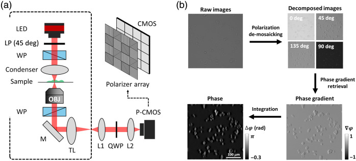
Label-free quantitative phase imaging can potentially measure cellular dynamics with minimal perturbation, motivating efforts to develop faster and more sensitive instrumentation. We characterize fast, single-shot quantitative phase gradient microscopy (ss-QPGM) that simultaneously acquires multiple polarization components required to reconstruct phase images. We integrate a computationally efficient least squares algorithm to provide real-time, video-rate imaging (up to ). The developed instrument was used to observe changes in cellular morphology and correlate these to molecular measures commonly obtained by staining.
We aim to characterize a fast approach to ss-QPGM and record morphological changes in single-cell phase images. We also correlate these with biochemical changes indicating cell death using concurrently acquired fluorescence images.
Here, we examine nutrient deprivation and anticancer drug-induced cell death in two different breast cell lines, , M2 and MCF7. Our approach involves in-line measurements of ss-QPGM and fluorescence imaging of the cells biochemically labeled for viability.
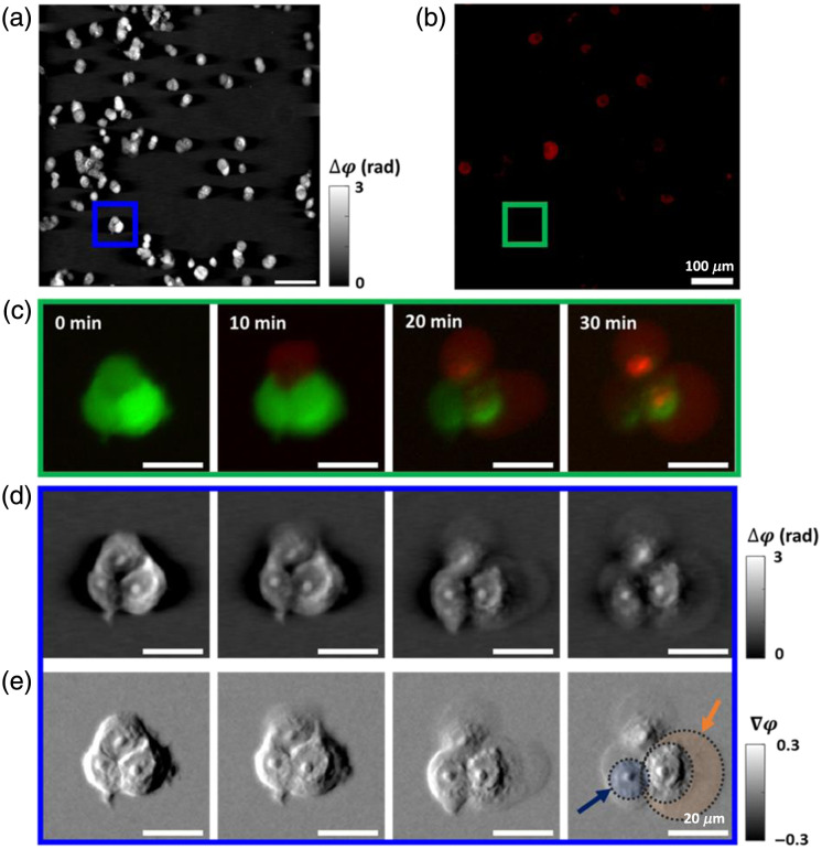
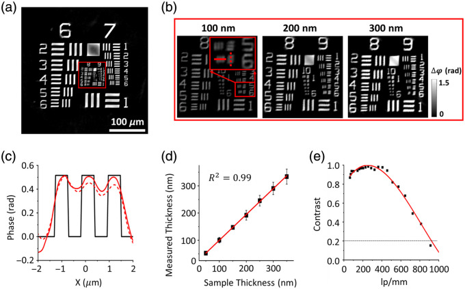
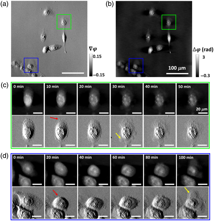
We validate the accuracy of the phase measurement using a USAF1951 pattern phase target. The ss-QPGM system resolves , and our analysis scheme accurately retrieves the phase with a high correlation coefficient ( ), as measured by calibrated sample thicknesses. Analyzing the contrast in phase, we estimate the spatial resolution achievable to be for this microscope. ss-QPGM time-lapse live-cell imaging reveals multiple intracellular and morphological changes during biochemically induced cell death. Inferences from co-registered images of quantitative phase and fluorescence suggest the possibility of necrosis, which agrees with previous findings.
Label-free ss-QPGM with high-temporal resolution and high spatial fidelity is demonstrated. Its application for monitoring dynamic changes in live cells offers promising prospects.
无标记定量相成像能够在最小程度的扰动下潜在地测量细胞动力学,这推动了人们致力于开发更快、更灵敏的仪器。我们对快速单次定量相梯度显微镜(ss-QPGM)进行了表征,该显微镜可同时获取重建相图像所需的多个偏振分量。我们集成了一种计算效率高的最小二乘法算法,以提供实时视频速率成像(高达 )。所开发的仪器用于观察细胞形态的变化,并将这些变化与通过染色通常获得的分子测量结果相关联。
我们旨在表征一种用于ss-QPGM的快速方法,并记录单细胞相图像中的形态变化。我们还使用同时获取的荧光图像将这些变化与指示细胞死亡的生化变化相关联。
在这里,我们研究了两种不同的乳腺癌细胞系 、M2和MCF7中营养剥夺和抗癌药物诱导的细胞死亡。我们的方法包括对ss-QPGM进行在线测量以及对经生化标记以检测活力的细胞进行荧光成像。
我们使用美国空军1951型图案相靶验证了相测量的准确性。ss-QPGM系统的分辨率为 ,并且我们的分析方案通过校准样品厚度测量,以高相关系数( )准确地检索到了相。通过分析相中的对比度,我们估计该显微镜可实现的空间分辨率为 。ss-QPGM延时活细胞成像揭示了生化诱导的细胞死亡过程中多种细胞内和形态学变化。定量相和荧光的配准图像推断表明存在坏死的可能性,这与先前的发现一致。
展示了具有高时间分辨率和高空间保真度的无标记ss-QPGM。其在监测活细胞动态变化方面的应用前景广阔。