Reddy C Kishor Kumar, Reddy Pulakurthi Anaghaa, Janapati Himaja, Assiri Basem, Shuaib Mohammed, Alam Shadab, Sheneamer Abdullah
Department of Computer Science and Engineering, Stanley College of Engineering and Technology for Women, Hyderabad, India.
Department of Computer Science, College of Engineering and Computer Science, Jazan University, Jazan, Saudi Arabia.
Front Oncol. 2024 Jul 18;14:1400341. doi: 10.3389/fonc.2024.1400341. eCollection 2024.
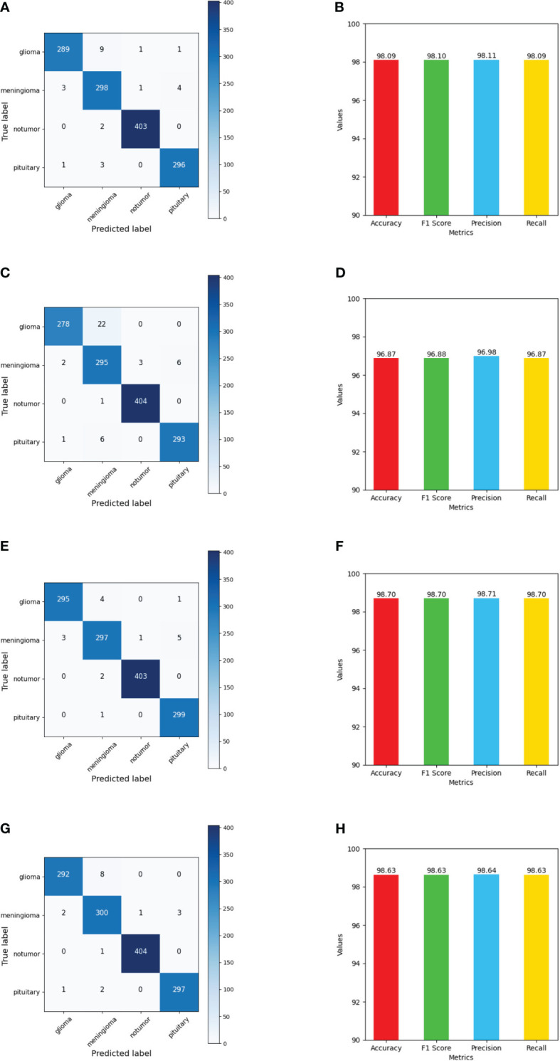
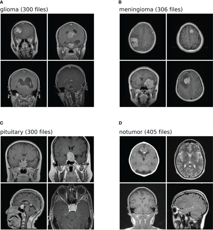
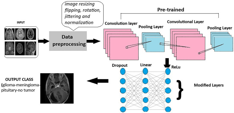
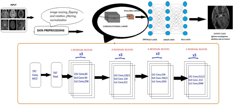
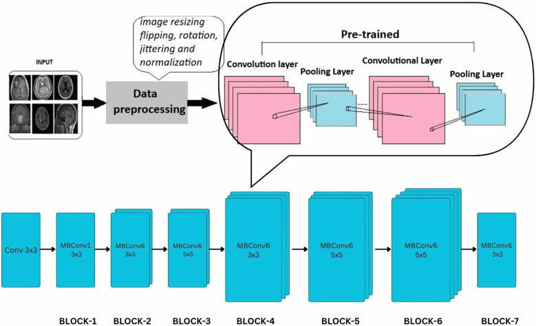
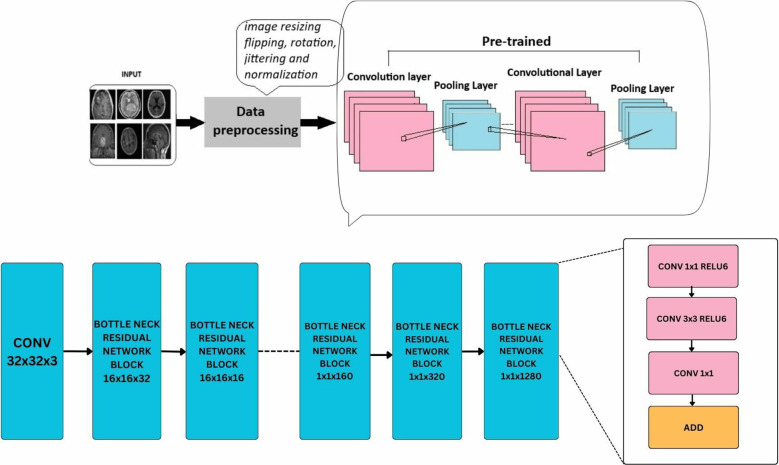
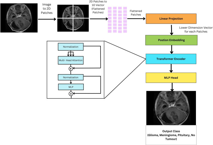
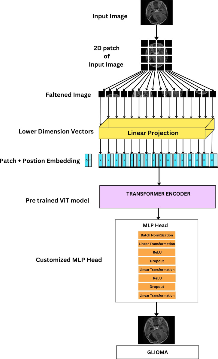
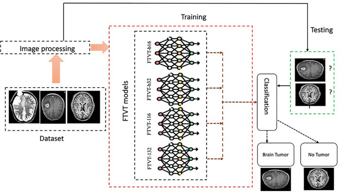
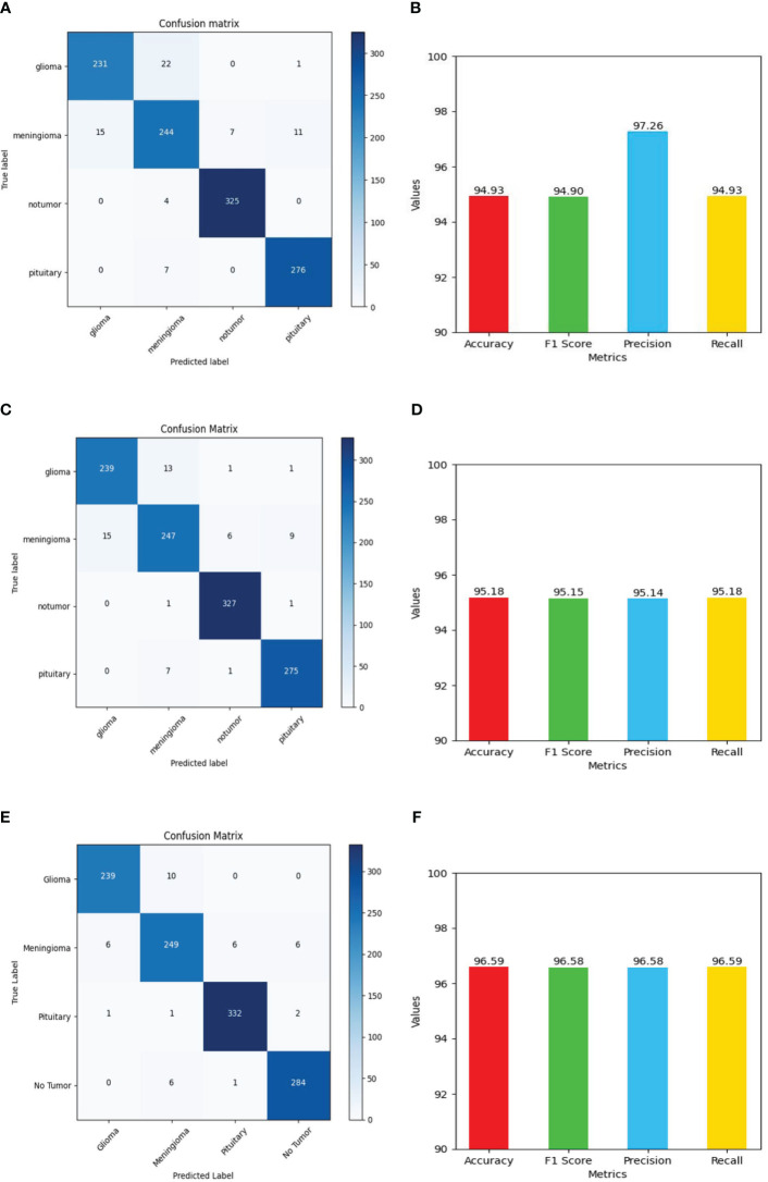
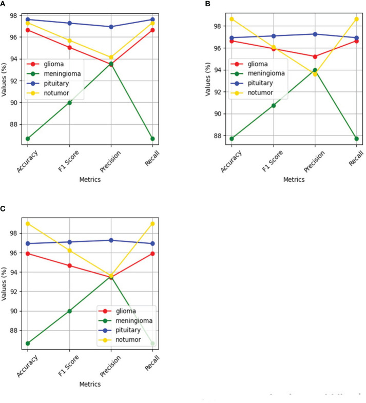
Brain tumors occur due to the expansion of abnormal cell tissues and can be malignant (cancerous) or benign (not cancerous). Numerous factors such as the position, size, and progression rate are considered while detecting and diagnosing brain tumors. Detecting brain tumors in their initial phases is vital for diagnosis where MRI (magnetic resonance imaging) scans play an important role. Over the years, deep learning models have been extensively used for medical image processing. The current study primarily investigates the novel Fine-Tuned Vision Transformer models (FTVTs)-FTVT-b16, FTVT-b32, FTVT-l16, FTVT-l32-for brain tumor classification, while also comparing them with other established deep learning models such as ResNet50, MobileNet-V2, and EfficientNet - B0. A dataset with 7,023 images (MRI scans) categorized into four different classes, namely, glioma, meningioma, pituitary, and no tumor are used for classification. Further, the study presents a comparative analysis of these models including their accuracies and other evaluation metrics including recall, precision, and F1-score across each class. The deep learning models ResNet-50, EfficientNet-B0, and MobileNet-V2 obtained an accuracy of 96.5%, 95.1%, and 94.9%, respectively. Among all the FTVT models, FTVT-l16 model achieved a remarkable accuracy of 98.70% whereas other FTVT models FTVT-b16, FTVT-b32, and FTVT-132 achieved an accuracy of 98.09%, 96.87%, 98.62%, respectively, hence proving the efficacy and robustness of FTVT's in medical image processing.
脑肿瘤是由于异常细胞组织的扩张而发生的,可分为恶性(癌性)或良性(非癌性)。在检测和诊断脑肿瘤时,会考虑许多因素,如位置、大小和进展速度。在诊断中,早期检测脑肿瘤至关重要,其中MRI(磁共振成像)扫描起着重要作用。多年来,深度学习模型已广泛应用于医学图像处理。当前的研究主要调查新型的微调视觉Transformer模型(FTVT)——FTVT-b16、FTVT-b32、FTVT-l16、FTVT-l32——用于脑肿瘤分类,同时还将它们与其他已建立的深度学习模型进行比较,如ResNet50、MobileNet-V2和EfficientNet - B0。一个包含7023张图像(MRI扫描)的数据集被分为四个不同类别,即神经胶质瘤、脑膜瘤、垂体瘤和无肿瘤,用于分类。此外,该研究对这些模型进行了比较分析,包括它们的准确率以及其他评估指标,包括每个类别的召回率、精确率和F1分数。深度学习模型ResNet-50、EfficientNet-B0和MobileNet-V2的准确率分别为96.5%、95.1%和94.9%。在所有FTVT模型中,FTVT-l16模型取得了98.70%的显著准确率,而其他FTVT模型FTVT-b16、FTVT-b32和FTVT-132的准确率分别为98.09%、96.87%、98.62%,从而证明了FTVT在医学图像处理中的有效性和稳健性。