• 文献检索
  • 文档翻译
  • 深度研究
  • 学术资讯
  • Suppr Zotero 插件Zotero 插件
  • 邀请有礼
  • 套餐&价格
  • 历史记录
应用&插件
Suppr Zotero 插件Zotero 插件浏览器插件Mac 客户端Windows 客户端微信小程序
定价
高级版会员购买积分包购买API积分包
服务
文献检索文档翻译深度研究API 文档MCP 服务
关于我们
关于 Suppr公司介绍联系我们用户协议隐私条款
关注我们

Suppr 超能文献

核心技术专利:CN118964589B侵权必究
粤ICP备2023148730 号-1Suppr @ 2026

文献检索

告别复杂PubMed语法,用中文像聊天一样搜索,搜遍4000万医学文献。AI智能推荐,让科研检索更轻松。

立即免费搜索

文件翻译

保留排版,准确专业,支持PDF/Word/PPT等文件格式,支持 12+语言互译。

免费翻译文档

深度研究

AI帮你快速写综述,25分钟生成高质量综述,智能提取关键信息,辅助科研写作。

立即免费体验

肌联蛋白通过微管和肌动蛋白以及细胞外基质主要支撑来调节心肌的被动僵硬。

Titin governs myocardial passive stiffness with major support from microtubules and actin and the extracellular matrix.

机构信息

Institute of Physiology II, University of Muenster, Muenster, Germany.

出版信息

Nat Cardiovasc Res. 2023 Nov;2(11):991-1002. doi: 10.1038/s44161-023-00348-1. Epub 2023 Oct 26.

DOI:10.1038/s44161-023-00348-1
PMID:39196092
原文链接:https://pmc.ncbi.nlm.nih.gov/articles/PMC11358001/
Abstract

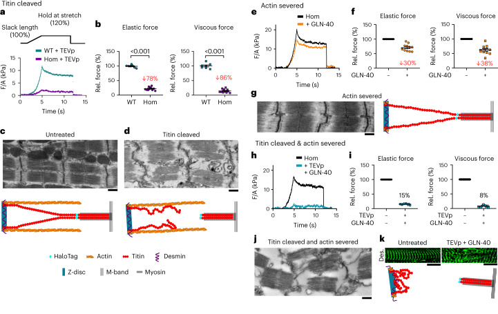
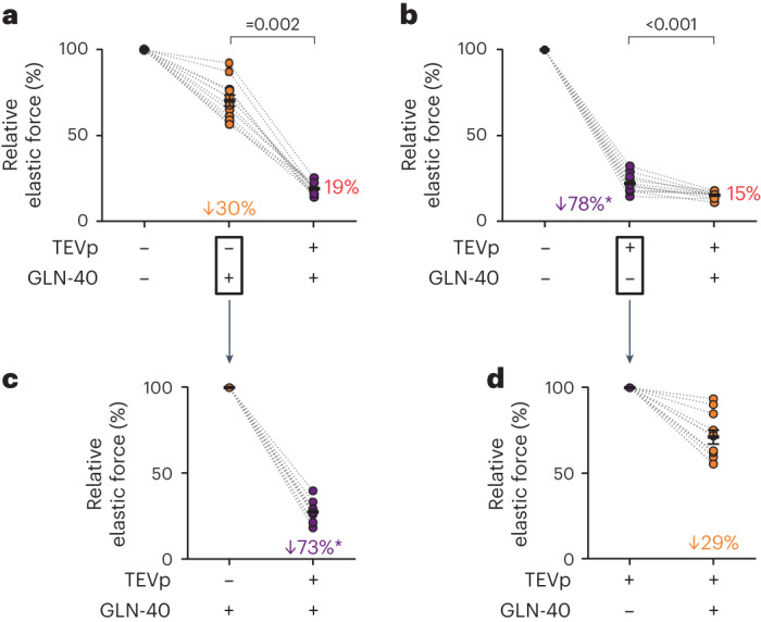
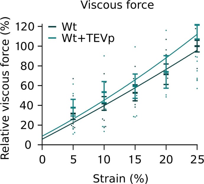
Myocardial passive stiffness is crucial for the heart's pump function and is determined by mechanical elements, including the extracellular matrix and cytoskeletal filaments; however, their individual contributions are controversially discussed and difficult to quantify. In this study, we targeted the cytoskeletal filaments in a mouse model, which enables the specific, acute and complete cleavage of the sarcomeric titin springs. We show in vitro that each cytoskeletal filament's stiffness contribution varies depending on whether the elastic or the viscous forces are considered and on strain level. Titin governs myocardial elastic forces, with the largest contribution provided at both low and high strain. Viscous force contributions are more uniformly distributed among the microtubules, titin and actin. The extracellular matrix contributes at high strain. The remaining forces after total target element disruption are likely derived from desmin filaments. Our findings answer longstanding questions about cardiac mechanical architecture and allow better targeting of passive myocardial stiffness in heart failure.

摘要

心肌被动僵硬度对心脏的泵功能至关重要,由机械元素决定,包括细胞外基质和细胞骨架丝;然而,它们各自的贡献存在争议,难以量化。在这项研究中,我们针对小鼠模型中的细胞骨架丝,实现了肌联蛋白弹簧的特异性、急性和完全切割。我们在体外表明,每条细胞骨架丝的刚度贡献取决于是考虑弹性力还是粘性力,以及应变水平。肌联蛋白控制心肌的弹性力,在低应变和高应变时都有最大的贡献。粘性力的贡献在微管、肌联蛋白和肌动蛋白之间更均匀地分布。细胞外基质在高应变时起作用。在完全破坏目标元素后剩余的力可能来自结蛋白丝。我们的发现回答了关于心脏机械结构的长期问题,并允许更好地针对心力衰竭中的心肌被动僵硬度。

https://cdn.ncbi.nlm.nih.gov/pmc/blobs/9021/11358001/e0783e4bd12c/44161_2023_348_Fig14_ESM.jpg
https://cdn.ncbi.nlm.nih.gov/pmc/blobs/9021/11358001/bf3f6d0c9229/44161_2023_348_Fig1_HTML.jpg
https://cdn.ncbi.nlm.nih.gov/pmc/blobs/9021/11358001/3a19e72fb60a/44161_2023_348_Fig2_HTML.jpg
https://cdn.ncbi.nlm.nih.gov/pmc/blobs/9021/11358001/804df04e40b3/44161_2023_348_Fig3_HTML.jpg
https://cdn.ncbi.nlm.nih.gov/pmc/blobs/9021/11358001/3b9d06b58051/44161_2023_348_Fig4_HTML.jpg
https://cdn.ncbi.nlm.nih.gov/pmc/blobs/9021/11358001/d1ced9f90f49/44161_2023_348_Fig5_HTML.jpg
https://cdn.ncbi.nlm.nih.gov/pmc/blobs/9021/11358001/f56949527b29/44161_2023_348_Fig6_HTML.jpg
https://cdn.ncbi.nlm.nih.gov/pmc/blobs/9021/11358001/203b4615b3c2/44161_2023_348_Fig7_HTML.jpg
https://cdn.ncbi.nlm.nih.gov/pmc/blobs/9021/11358001/7d58bb18b838/44161_2023_348_Fig8_ESM.jpg
https://cdn.ncbi.nlm.nih.gov/pmc/blobs/9021/11358001/e8cf17d06df3/44161_2023_348_Fig9_ESM.jpg
https://cdn.ncbi.nlm.nih.gov/pmc/blobs/9021/11358001/0e9e31381812/44161_2023_348_Fig10_ESM.jpg
https://cdn.ncbi.nlm.nih.gov/pmc/blobs/9021/11358001/da4f635ad2c7/44161_2023_348_Fig11_ESM.jpg
https://cdn.ncbi.nlm.nih.gov/pmc/blobs/9021/11358001/b2da1c3a630d/44161_2023_348_Fig12_ESM.jpg
https://cdn.ncbi.nlm.nih.gov/pmc/blobs/9021/11358001/cf05f2b5e07b/44161_2023_348_Fig13_ESM.jpg
https://cdn.ncbi.nlm.nih.gov/pmc/blobs/9021/11358001/e0783e4bd12c/44161_2023_348_Fig14_ESM.jpg

相似文献

1
Titin governs myocardial passive stiffness with major support from microtubules and actin and the extracellular matrix.肌联蛋白通过微管和肌动蛋白以及细胞外基质主要支撑来调节心肌的被动僵硬。
Nat Cardiovasc Res. 2023 Nov;2(11):991-1002. doi: 10.1038/s44161-023-00348-1. Epub 2023 Oct 26.
2
Titin takes centerstage among cytoskeletal contributions to myocardial passive stiffness.肌联蛋白在细胞骨架对心肌被动僵硬的贡献中占据中心地位。
Cytoskeleton (Hoboken). 2024 Feb-Mar;81(2-3):184-187. doi: 10.1002/cm.21827. Epub 2023 Dec 29.
3
Cardiac titin: molecular basis of elasticity and cellular contribution to elastic and viscous stiffness components in myocardium.心肌肌联蛋白:弹性的分子基础以及细胞对心肌弹性和粘性硬度成分的贡献
J Muscle Res Cell Motil. 2002;23(5-6):483-97. doi: 10.1023/a:1023462507254.
4
Actin-titin interaction in cardiac myofibrils: probing a physiological role.心肌肌原纤维中肌动蛋白与肌联蛋白的相互作用:探索其生理作用
Biophys J. 1997 Aug;73(2):905-19. doi: 10.1016/S0006-3495(97)78123-2.
5
Titin-isoform dependence of titin-actin interaction and its regulation by S100A1/Ca2+ in skinned myocardium.在去垢剂处理的心肌中,肌联蛋白与肌动蛋白相互作用对肌联蛋白异构体的依赖性及其受S100A1/Ca2+的调节
J Biomed Biotechnol. 2010;2010:727239. doi: 10.1155/2010/727239. Epub 2010 Apr 14.
6
Passive tension in cardiac muscle: contribution of collagen, titin, microtubules, and intermediate filaments.心肌中的被动张力:胶原蛋白、肌联蛋白、微管和中间丝的作用
Biophys J. 1995 Mar;68(3):1027-44. doi: 10.1016/S0006-3495(95)80278-X.
7
Hyperphosphorylation of mouse cardiac titin contributes to transverse aortic constriction-induced diastolic dysfunction.肌联蛋白在小鼠心脏中的过度磷酸化导致了主动脉缩窄引起的舒张功能障碍。
Circ Res. 2011 Sep 30;109(8):858-66. doi: 10.1161/CIRCRESAHA.111.246819. Epub 2011 Aug 11.
8
Interaction between PEVK-titin and actin filaments: origin of a viscous force component in cardiac myofibrils.肌联蛋白富含脯氨酸(PEVK)区域与肌动蛋白丝之间的相互作用:心肌肌原纤维中粘性力成分的起源
Circ Res. 2001 Nov 9;89(10):874-81. doi: 10.1161/hh2201.099453.
9
Titin elasticity and mechanism of passive force development in rat cardiac myocytes probed by thin-filament extraction.通过细肌丝提取探究大鼠心肌细胞中肌联蛋白弹性及被动力产生机制
Biophys J. 1997 Oct;73(4):2043-53. doi: 10.1016/S0006-3495(97)78234-1.
10
Protein kinase-A phosphorylates titin in human heart muscle and reduces myofibrillar passive tension.蛋白激酶A使人类心肌中的肌联蛋白磷酸化,并降低肌原纤维的被动张力。
J Muscle Res Cell Motil. 2006;27(5-7):435-44. doi: 10.1007/s10974-006-9090-5. Epub 2006 Aug 4.

引用本文的文献

1
Persistent Fibrosis and Left Ventricular Chamber Stiffening Despite Cessation of Repetitive Pressure Overload in Swine.尽管猪的重复性压力超负荷已停止,但仍存在持续性纤维化和左心室腔硬化。
JACC Basic Transl Sci. 2025 Jun;10(6):844-859. doi: 10.1016/j.jacbts.2025.02.009. Epub 2025 Apr 30.
2
Direction-dependent contributions of cardiac myofilament networks to myocardial passive stiffness reveal a major disparity for titin.心肌肌丝网络对心肌被动僵硬度的方向依赖性贡献揭示了肌联蛋白的一个主要差异。
Basic Res Cardiol. 2025 Jun 13. doi: 10.1007/s00395-025-01119-8.
3
Vasohibins in Health and Disease: From Angiogenesis to Tumorigenesis, Multiorgan Dysfunction, and Brain-Heart Remodeling.

本文引用的文献

1
A guide for assessment of myocardial stiffness in health and disease.健康与疾病状态下心肌僵硬度评估指南。
Nat Cardiovasc Res. 2022 Jan;1(1):8-22. doi: 10.1038/s44161-021-00007-3. Epub 2022 Jan 12.
2
Titin force in muscle cells alters lattice order, thick and thin filament protein formation.肌球蛋白在肌肉细胞中的作用力改变了晶格顺序,厚和薄的丝蛋白的形成。
Proc Natl Acad Sci U S A. 2022 Nov 29;119(48):e2209441119. doi: 10.1073/pnas.2209441119. Epub 2022 Nov 21.
3
Desmin intermediate filaments and tubulin detyrosination stabilize growing microtubules in the cardiomyocyte.
健康与疾病中的血管抑制素:从血管生成到肿瘤发生、多器官功能障碍及脑心重塑
Cells. 2025 May 23;14(11):767. doi: 10.3390/cells14110767.
4
Mechanically knocking out titin reveals protein tension loss as a trigger of muscle disease.机械敲除肌联蛋白揭示蛋白质张力丧失是肌肉疾病的触发因素。
Nat Biomed Eng. 2025 Jun 5. doi: 10.1038/s41551-025-01403-x.
5
Mechanism-based myofilament manipulation to treat diastolic dysfunction in HFpEF.基于机制的肌丝调控以治疗射血分数保留的心力衰竭中的舒张功能障碍。
Front Physiol. 2024 Dec 12;15:1512550. doi: 10.3389/fphys.2024.1512550. eCollection 2024.
6
Alternative Splicing in the Heart: The Therapeutic Potential of Regulating the Regulators.心脏中的可变剪接:调控调控因子的治疗潜力
Int J Mol Sci. 2024 Dec 4;25(23):13023. doi: 10.3390/ijms252313023.
7
Mechanical loading reveals an intrinsic cardiomyocyte stiffness contribution to diastolic dysfunction in murine cardiometabolic disease.机械负荷揭示了心肌细胞固有僵硬度对小鼠心脏代谢疾病舒张功能障碍的影响。
J Physiol. 2024 Dec;602(24):6705-6727. doi: 10.1113/JP286437. Epub 2024 Dec 4.
8
Titin: roles in cardiac function and diseases.肌联蛋白:在心脏功能和疾病中的作用
Front Physiol. 2024 Apr 10;15:1385821. doi: 10.3389/fphys.2024.1385821. eCollection 2024.
9
Microtubule destabilization with colchicine increases the work output of myocardial slices.秋水仙碱使微管解聚可增加心肌切片的功输出。
J Mol Cell Cardiol Plus. 2024 Mar;7. doi: 10.1016/j.jmccpl.2024.100066. Epub 2024 Feb 29.
10
Titin-Based Force Modulates Cardiac Thick and Thin Filaments.基于肌联蛋白的力调节心脏粗细肌丝。
Circ Res. 2024 Apr 12;134(8):1026-1028. doi: 10.1161/CIRCRESAHA.123.323988. Epub 2024 Mar 14.
结蛋白中间丝和微管去酪氨酸化稳定心肌细胞中的生长微管。
Basic Res Cardiol. 2022 Nov 3;117(1):53. doi: 10.1007/s00395-022-00962-3.
4
Transcriptional, Post-Transcriptional, and Post-Translational Mechanisms Rewrite the Tubulin Code During Cardiac Hypertrophy and Failure.转录、转录后和翻译后机制在心脏肥大和衰竭过程中重写微管蛋白密码。
Front Cell Dev Biol. 2022 Apr 1;10:837486. doi: 10.3389/fcell.2022.837486. eCollection 2022.
5
Therapeutic potential of colchicine in cardiovascular medicine: a pharmacological review.秋水仙碱在心血管医学中的治疗潜力:药理学评价。
Acta Pharmacol Sin. 2022 Sep;43(9):2173-2190. doi: 10.1038/s41401-021-00835-w. Epub 2022 Jan 19.
6
Functional and structural differences between skinned and intact muscle preparations.去皮和完整肌肉标本的功能和结构差异。
J Gen Physiol. 2022 Feb 7;154(2). doi: 10.1085/jgp.202112990. Epub 2022 Jan 19.
7
Truncated titin proteins and titin haploinsufficiency are targets for functional recovery in human cardiomyopathy due to mutations.截短的肌联蛋白蛋白和肌联蛋白部分功能缺失是由于基因突变导致的人类心肌病功能恢复的靶点。
Sci Transl Med. 2021 Nov 3;13(618):eabd3079. doi: 10.1126/scitranslmed.abd3079.
8
Titin (TTN): from molecule to modifications, mechanics, and medical significance.肌联蛋白(TTN):从分子到修饰、力学和医学意义。
Cardiovasc Res. 2022 Nov 10;118(14):2903-2918. doi: 10.1093/cvr/cvab328.
9
Cardiomyocyte Microtubules: Control of Mechanics, Transport, and Remodeling.心肌细胞微管:力学、运输和重构的控制。
Annu Rev Physiol. 2022 Feb 10;84:257-283. doi: 10.1146/annurev-physiol-062421-040656. Epub 2021 Oct 6.
10
Mechanisms of Frank-Starling law of the heart and stretch activation in striated muscles may have a common molecular origin.心肌的 Frank-Starling 定律和横纹肌牵张激活的机制可能具有共同的分子起源。
J Muscle Res Cell Motil. 2021 Jun;42(2):355-366. doi: 10.1007/s10974-020-09595-2. Epub 2021 Feb 11.