• 文献检索
  • 文档翻译
  • 深度研究
  • 学术资讯
  • Suppr Zotero 插件Zotero 插件
  • 邀请有礼
  • 套餐&价格
  • 历史记录
应用&插件
Suppr Zotero 插件Zotero 插件浏览器插件Mac 客户端Windows 客户端微信小程序
定价
高级版会员购买积分包购买API积分包
服务
文献检索文档翻译深度研究API 文档MCP 服务
关于我们
关于 Suppr公司介绍联系我们用户协议隐私条款
关注我们

Suppr 超能文献

核心技术专利:CN118964589B侵权必究
粤ICP备2023148730 号-1Suppr @ 2026

文献检索

告别复杂PubMed语法,用中文像聊天一样搜索,搜遍4000万医学文献。AI智能推荐,让科研检索更轻松。

立即免费搜索

文件翻译

保留排版,准确专业,支持PDF/Word/PPT等文件格式,支持 12+语言互译。

免费翻译文档

深度研究

AI帮你快速写综述,25分钟生成高质量综述,智能提取关键信息,辅助科研写作。

立即免费体验

用于改善早期浸润性肺腺癌诊断的人工智能驱动的组织学模式识别

: artificial intelligence-driven histologic pattern recognition for improved diagnosis of early-stage invasive lung adenocarcinoma.

作者信息

Huang Haoda, Yan Zeping, Li Bingliang, Lu Weixiang, He Ping, Fan Lei, Wu Xiaowei, Liang Hengrui, He Jianxing

机构信息

Nanfang Hospital, Southern Medical University, Guangzhou, China.

Department of Thoracic Surgery, the First Affiliated Hospital of Guangzhou Medical University, Guangzhou, China.

出版信息

Transl Lung Cancer Res. 2024 Aug 31;13(8):1816-1827. doi: 10.21037/tlcr-24-258. Epub 2024 Aug 26.

DOI:10.21037/tlcr-24-258
PMID:39263012
原文链接:https://pmc.ncbi.nlm.nih.gov/articles/PMC11384487/
Abstract

BACKGROUND

Early-stage invasive lung adenocarcinoma (ADC) characterized by a predominant micropapillary or solid pattern exhibit an elevated risk of recurrence following sub-lobar resection, thus determining histological subtype of early-stage invasive ADC prior surgery is important for formulating lobectomy or sub-lobar resection. This study aims to develop a deep learning algorithm and assess its clinical capability in distinguishing high-risk or low-risk histologic patterns in early-stage invasive ADC based on preoperative computed tomography (CT) scans.

METHODS

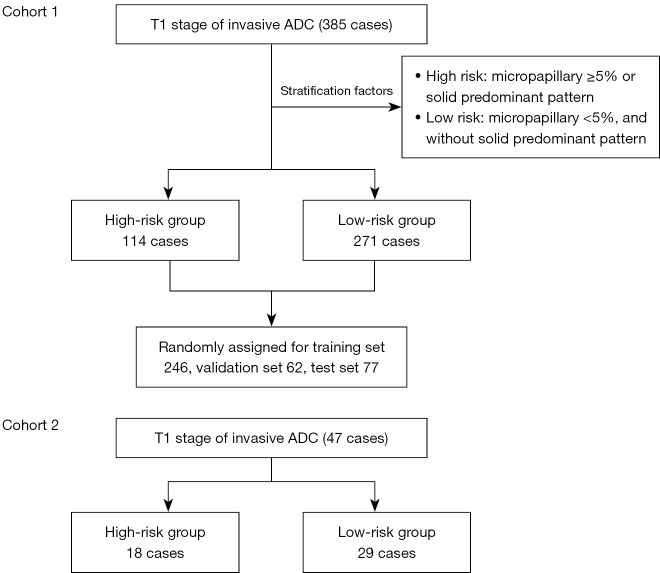
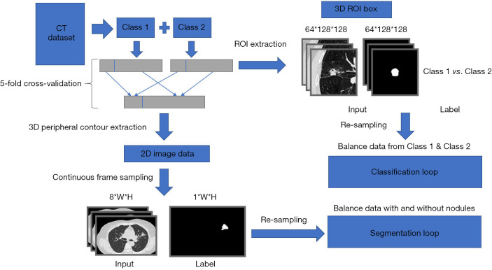
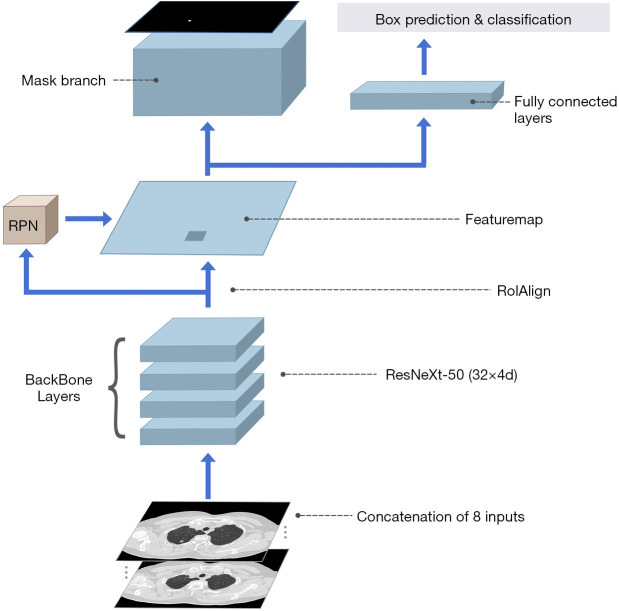
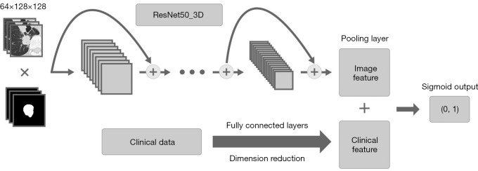
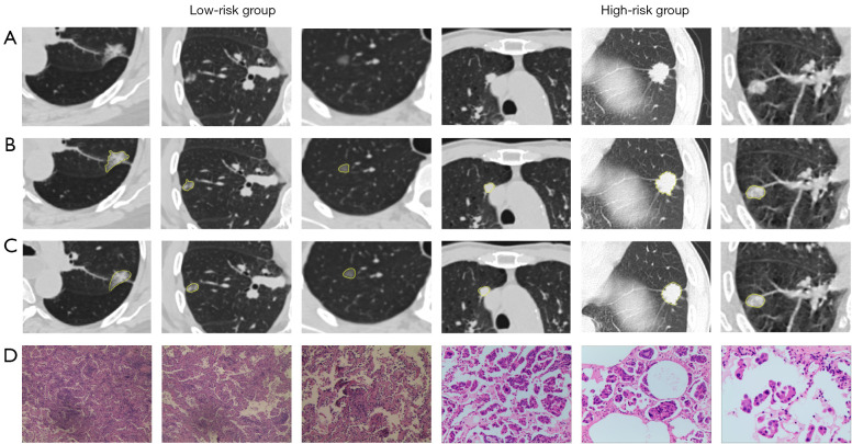
Two retrospective cohorts were included: development cohort 1 and external test cohort 2, comprising patients diagnosed with T1 stage invasive ADC. Electronic medical records and CT scans of all patients were documented. Patients were stratified into two risk groups. High-risk group: comprising cases with a micropapillary component ≥5% or a predominant solid pattern. Low-risk group: encompassing cases with a micropapillary component <5% and an absence of a predominant solid pattern. The overall segmentation model was modified based on Mask Region-based Convolutional Neural Network (Mask-RCNN), and Residual Network 50 (ResNet50)_3D was employed for image classification.

RESULTS

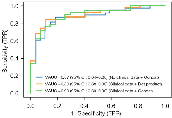
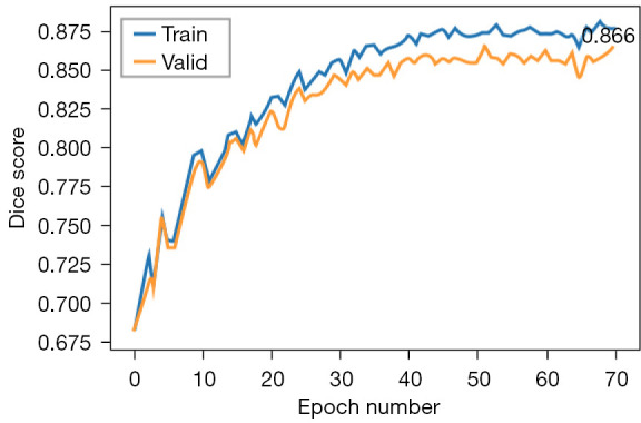
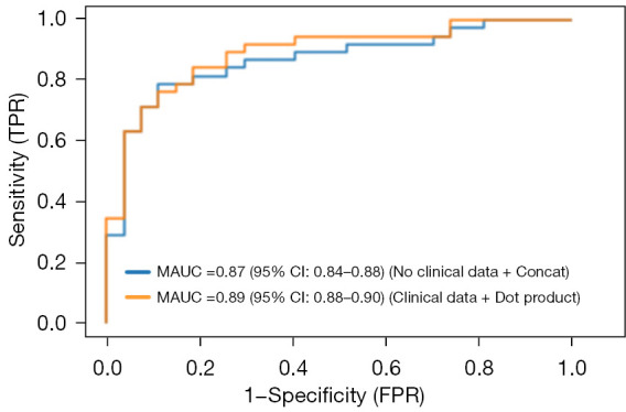
A total of 432 patients participated in this study, with 385 cases in cohort 1 and 47 cases in cohort 2. The fine-outline results produced by the auto-segmentation model exhibited a high level of agreement with manual segmentation by human experts, yielding a mean dice coefficient of 0.86 [95% confidence interval (CI): 0.85-0.87] in cohort 1 and 0.84 (95% CI: 0.82-0.85) in cohort 2. Furthermore, the deep learning model effectively differentiated the high-risk group from the low-risk group, achieving an area under the curve (AUC) of 0.89 (95% CI: 0.88-0.90) in cohort 1. In the external validation conducted in cohort 2, the deep learning model displayed an AUC of 0.87 (95% CI: 0.84-0.88) in distinguishing the high-risk group from the low-risk group. The average diagnostic time was 16.00±3.2 seconds, with an accuracy of 0.82 (95% CI: 0.81-0.83).

CONCLUSIONS

We have developed a deep learning algorithm, , for the automated segmentation of pulmonary nodules and prediction of high-risk histological patterns in early-stage lung ADC based on CT scans.

摘要

背景

以微乳头或实性为主型为特征的早期浸润性肺腺癌(ADC)在亚肺叶切除术后复发风险升高,因此在手术前确定早期浸润性ADC的组织学亚型对于制定肺叶切除术或亚肺叶切除术很重要。本研究旨在开发一种深度学习算法,并评估其基于术前计算机断层扫描(CT)区分早期浸润性ADC中高风险或低风险组织学模式的临床能力。

方法

纳入两个回顾性队列:开发队列1和外部测试队列2,包括诊断为T1期浸润性ADC的患者。记录所有患者的电子病历和CT扫描。患者被分为两个风险组。高风险组:包括微乳头成分≥5%或实性为主型的病例。低风险组:包括微乳头成分<5%且无实性为主型的病例。基于掩膜区域卷积神经网络(Mask-RCNN)修改整体分割模型,并采用残差网络50(ResNet50)_3D进行图像分类。

结果

共有432例患者参与本研究,队列1中有385例,队列2中有47例。自动分割模型产生的精细轮廓结果与人类专家的手动分割高度一致,队列1中的平均骰子系数为0.86 [95%置信区间(CI):0.85 - 0.87],队列2中为0.84(95% CI:0.82 - 0.85)。此外,深度学习模型有效地将高风险组与低风险组区分开来,队列1中的曲线下面积(AUC)为0.89(95% CI:0.88 - 0.90)。在队列2中进行的外部验证中,深度学习模型在区分高风险组与低风险组时的AUC为0.87(95% CI:0.84 - 0.88)。平均诊断时间为16.00±3.2秒,准确率为0.82(95% CI:0.81 - 0.83)。

结论

我们开发了一种深度学习算法,用于基于CT扫描对肺结节进行自动分割,并预测早期肺ADC中的高风险组织学模式。

https://cdn.ncbi.nlm.nih.gov/pmc/blobs/09bc/11384487/9b3b08533e64/tlcr-13-08-1816-f8.jpg
https://cdn.ncbi.nlm.nih.gov/pmc/blobs/09bc/11384487/001ca3769c31/tlcr-13-08-1816-f1.jpg
https://cdn.ncbi.nlm.nih.gov/pmc/blobs/09bc/11384487/ca93907234cc/tlcr-13-08-1816-f2.jpg
https://cdn.ncbi.nlm.nih.gov/pmc/blobs/09bc/11384487/fe3ec7693c4c/tlcr-13-08-1816-f3.jpg
https://cdn.ncbi.nlm.nih.gov/pmc/blobs/09bc/11384487/78245b6c4b24/tlcr-13-08-1816-f4.jpg
https://cdn.ncbi.nlm.nih.gov/pmc/blobs/09bc/11384487/4d5684f927b0/tlcr-13-08-1816-f5.jpg
https://cdn.ncbi.nlm.nih.gov/pmc/blobs/09bc/11384487/8cf71cdb9c56/tlcr-13-08-1816-f6.jpg
https://cdn.ncbi.nlm.nih.gov/pmc/blobs/09bc/11384487/6e8362ecdb6d/tlcr-13-08-1816-f7.jpg
https://cdn.ncbi.nlm.nih.gov/pmc/blobs/09bc/11384487/9b3b08533e64/tlcr-13-08-1816-f8.jpg

相似文献

1
: artificial intelligence-driven histologic pattern recognition for improved diagnosis of early-stage invasive lung adenocarcinoma.用于改善早期浸润性肺腺癌诊断的人工智能驱动的组织学模式识别
Transl Lung Cancer Res. 2024 Aug 31;13(8):1816-1827. doi: 10.21037/tlcr-24-258. Epub 2024 Aug 26.
2
Development and validation of a deep learning-based framework for automated lung CT segmentation and acute respiratory distress syndrome prediction: a multicenter cohort study.基于深度学习的自动肺CT分割和急性呼吸窘迫综合征预测框架的开发与验证:一项多中心队列研究
EClinicalMedicine. 2024 Jul 26;75:102772. doi: 10.1016/j.eclinm.2024.102772. eCollection 2024 Sep.
3
CT-Based Deep Learning Model for Invasiveness Classification and Micropapillary Pattern Prediction Within Lung Adenocarcinoma.基于CT的深度学习模型用于肺腺癌侵袭性分类和微乳头模式预测
Front Oncol. 2020 Jul 22;10:1186. doi: 10.3389/fonc.2020.01186. eCollection 2020.
4
[The correlation between metabolic parameters in (18)F-FDG PET-CT and solid and micropapillary histological subtypes in lung adenocarcinoma].[(18)F-FDG PET-CT代谢参数与肺腺癌实性及微乳头组织学亚型之间的相关性]
Zhonghua Zhong Liu Za Zhi. 2022 Jun 23;44(6):555-561. doi: 10.3760/cma.j.cn112152-20200804-00710.
5
Deep learning-based diagnosis of histopathological patterns for invasive non-mucinous lung adenocarcinoma using semantic segmentation.基于深度学习的浸润性非黏液肺腺癌组织病理模式的语义分割诊断。
BMJ Open. 2023 Jul 25;13(7):e069181. doi: 10.1136/bmjopen-2022-069181.
6
Computed Tomography Image under Convolutional Neural Network Deep Learning Algorithm in Pulmonary Nodule Detection and Lung Function Examination.卷积神经网络深度学习算法在肺结节检测和肺功能检查中的计算机断层扫描图像。
J Healthc Eng. 2021 Oct 22;2021:3417285. doi: 10.1155/2021/3417285. eCollection 2021.
7
Computed tomography-based 3D convolutional neural network deep learning model for predicting micropapillary or solid growth pattern of invasive lung adenocarcinoma.基于计算机断层扫描的三维卷积神经网络深度学习模型预测浸润性肺腺癌的微乳头状或实性生长模式。
Radiol Med. 2024 May;129(5):776-784. doi: 10.1007/s11547-024-01800-3. Epub 2024 Mar 21.
8
Design of lung nodules segmentation and recognition algorithm based on deep learning.基于深度学习的肺结节分割与识别算法设计。
BMC Bioinformatics. 2021 Nov 8;22(Suppl 5):314. doi: 10.1186/s12859-021-04234-0.
9
Clinical, Imaging, and Pathological-Molecular Characteristics Associated with Stage IA Invasive Lung Adenocarcinoma Recurrence After Sub-lobar Resection.与肺叶下切除术后IA期浸润性肺腺癌复发相关的临床、影像学及病理分子特征
Acad Radiol. 2025 Jan;32(1):450-459. doi: 10.1016/j.acra.2024.07.003. Epub 2024 Jul 22.
10
Artificial intelligence-based recurrence prediction outperforms classical histopathological methods in pulmonary adenocarcinoma biopsies.基于人工智能的复发预测在肺腺癌活检中优于传统组织病理学方法。
Lung Cancer. 2023 Dec;186:107413. doi: 10.1016/j.lungcan.2023.107413. Epub 2023 Nov 4.

引用本文的文献

1
Effectiveness of AI for Enhancing Computed Tomography Image Quality and Radiation Protection in Radiology: Systematic Review and Meta-Analysis.人工智能在提高放射学中计算机断层扫描图像质量和辐射防护方面的有效性:系统评价与荟萃分析
J Med Internet Res. 2025 Feb 27;27:e66622. doi: 10.2196/66622.

本文引用的文献

1
Minor (≤ 10%) Ground-Glass Opacity Component in Clinical Stage I Non-Small Cell Lung Cancer: Associations With Pathologic Characteristics and Clinical Outcomes.临床Ⅰ期非小细胞肺癌中≤10%磨玻璃密度成分:与病理特征和临床结局的关系。
AJR Am J Roentgenol. 2024 Aug;223(2):e2431283. doi: 10.2214/AJR.24.31283. Epub 2024 May 29.
2
Prediction of the pathological subtypes by intraoperative frozen section for patients with cT1N0M0 invasive lung adenocarcinoma (ECTOP-1015): a prospective multicenter study.cT1N0M0 期浸润性肺腺癌患者术中冰冻切片预测病理亚型(ECTOP-1015):一项前瞻性多中心研究。
Int J Surg. 2024 Sep 1;110(9):5444-5451. doi: 10.1097/JS9.0000000000001667.
3
Sub-lobar resection versus lobectomy for challenging intraoperative frozen sections in lung adenocarcinoma within 3 cm.
对于3厘米以内肺腺癌术中冰冻切片有挑战性的情况,肺段切除术与肺叶切除术的比较
Asian J Surg. 2024 Dec;47(12):5113-5117. doi: 10.1016/j.asjsur.2024.05.002. Epub 2024 May 17.
4
A combination of radiomic features, clinic characteristics, and serum tumor biomarkers to predict the possibility of the micropapillary/solid component of lung adenocarcinoma.联合放射组学特征、临床特征和血清肿瘤标志物预测肺腺癌微乳头/实性成分的可能性。
Ther Adv Respir Dis. 2024 Jan-Dec;18:17534666241249168. doi: 10.1177/17534666241249168.
5
Computed tomography-based 3D convolutional neural network deep learning model for predicting micropapillary or solid growth pattern of invasive lung adenocarcinoma.基于计算机断层扫描的三维卷积神经网络深度学习模型预测浸润性肺腺癌的微乳头状或实性生长模式。
Radiol Med. 2024 May;129(5):776-784. doi: 10.1007/s11547-024-01800-3. Epub 2024 Mar 21.
6
The presence of micropapillary and/or solid subtypes is an independent prognostic factor for patients undergoing curative resection for stage I lung adenocarcinoma with ground-glass opacity.微乳头和/或实体亚型的存在是接受I期磨玻璃影肺腺癌根治性切除患者的独立预后因素。
Transl Lung Cancer Res. 2024 Feb 29;13(2):256-268. doi: 10.21037/tlcr-23-736. Epub 2024 Feb 20.
7
AI-guided histopathology predicts brain metastasis in lung cancer patients.人工智能引导的组织病理学预测肺癌患者的脑转移。
J Pathol. 2024 May;263(1):89-98. doi: 10.1002/path.6263. Epub 2024 Mar 4.
8
Micropapillary and solid components as high-grade patterns in IASLC grading system of lung adenocarcinoma: Clinical implications and management.IASLC 肺腺癌分级系统中微乳头和实性成分作为高级别模式:临床意义和管理。
Lung Cancer. 2024 Jan;187:107445. doi: 10.1016/j.lungcan.2023.107445. Epub 2023 Dec 16.
9
Radiologic Identification of Pathologic Tumor Invasion in Patients With Lung Adenocarcinoma.肺腺癌患者病理性肿瘤侵袭的放射学鉴定。
JAMA Netw Open. 2023 Oct 2;6(10):e2337889. doi: 10.1001/jamanetworkopen.2023.37889.
10
A Shift in Paradigm: Selective Lymph Node Dissection for Minimizing Oversurgery in Early Stage Lung Cancer.范式转变:选择性淋巴结清扫术以最小化早期肺癌过度手术
J Thorac Oncol. 2024 Jan;19(1):25-35. doi: 10.1016/j.jtho.2023.09.1443. Epub 2023 Sep 23.