Department of Biomedical Engineering, Tianjin Key Laboratory of Biomedical Detecting Techniques and Instruments, Tianjin University, Tianjin, China.
Department of Radiotherapy, Tianjin Medical University Cancer Institute and Hospital, National Clinical Research Center for Cancer, Tianjin Key Laboratory of Cancer Prevention and Therapy, Tianjin, China.
BMC Bioinformatics. 2021 Nov 8;22(Suppl 5):314. doi: 10.1186/s12859-021-04234-0.
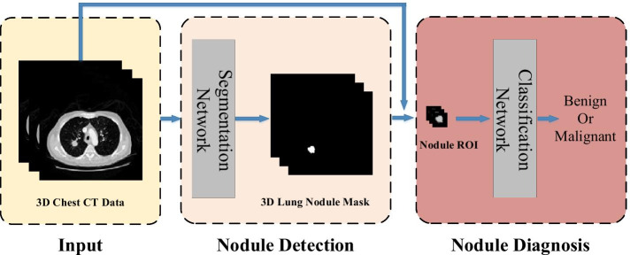
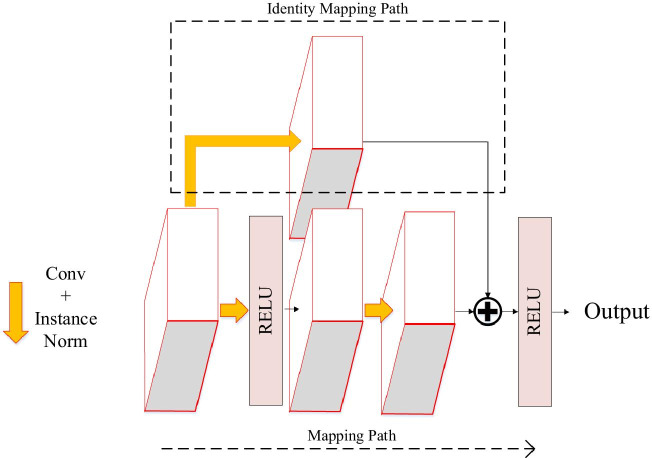
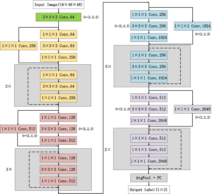
Accurate segmentation and recognition algorithm of lung nodules has great important value of reference for early diagnosis of lung cancer. An algorithm is proposed for 3D CT sequence images in this paper based on 3D Res U-Net segmentation network and 3D ResNet50 classification network. The common convolutional layers in encoding and decoding paths of U-Net are replaced by residual units while the loss function is changed to Dice loss after using cross entropy loss to accelerate network convergence. Since the lung nodules are small and rich in 3D information, the ResNet50 is improved by replacing the 2D convolutional layers with 3D convolutional layers and reducing the sizes of some convolution kernels, 3D ResNet50 network is obtained for the diagnosis of benign and malignant lung nodules.
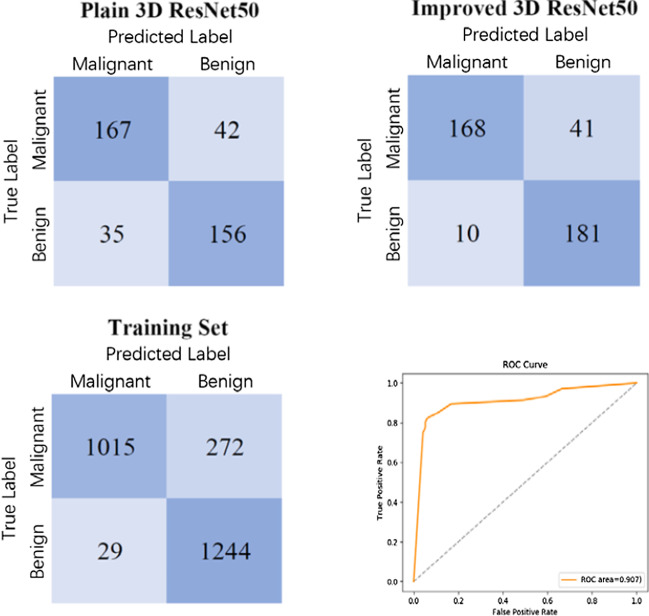
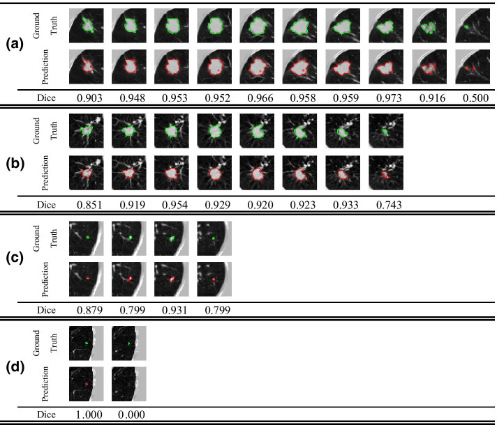
3D Res U-Net was trained and tested on 1044 CT subcases in the LIDC-IDRI database. The segmentation result shows that the Dice coefficient of 3D Res U-Net is above 0.8 for the segmentation of lung nodules larger than 10 mm in diameter. 3D ResNet50 was trained and tested on 2960 lung nodules in the LIDC-IDRI database. The classification result shows that the diagnostic accuracy of 3D ResNet50 is 87.3% and AUC is 0.907.
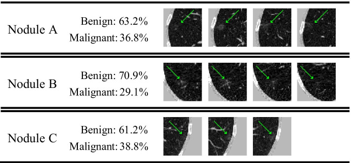
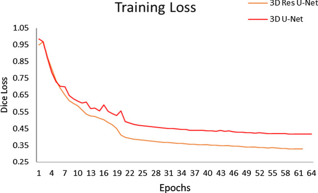
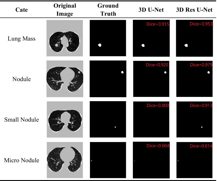
The 3D Res U-Net module improves segmentation performance significantly with the comparison of 3D U-Net model based on residual learning mechanism. 3D Res U-Net can identify small nodules more effectively and improve its segmentation accuracy for large nodules. Compared with the original network, the classification performance of 3D ResNet50 is significantly improved, especially for small benign nodules.
肺结节的准确分割和识别算法对肺癌的早期诊断具有重要的参考价值。本文提出了一种基于 3D Res U-Net 分割网络和 3D ResNet50 分类网络的 3D CT 序列图像算法。在使用交叉熵损失加速网络收敛后,将 U-Net 编码和解码路径中的常见卷积层替换为残差单元,同时将损失函数更改为 Dice 损失。由于肺结节体积小且富含 3D 信息,因此通过用 3D 卷积层替换 ResNet50 中的 2D 卷积层并减小一些卷积核的大小,从而得到用于诊断良性和恶性肺结节的 3D ResNet50 网络。
在 LIDC-IDRI 数据库的 1044 个 CT 子例中对 3D Res U-Net 进行了训练和测试。分割结果表明,对于直径大于 10mm 的肺结节,3D Res U-Net 的 Dice 系数大于 0.8。在 LIDC-IDRI 数据库的 2960 个肺结节上对 3D ResNet50 进行了训练和测试。分类结果表明,3D ResNet50 的诊断准确率为 87.3%,AUC 为 0.907。
与基于残差学习机制的 3D U-Net 模型相比,3D Res U-Net 模块显著提高了分割性能。3D Res U-Net 可以更有效地识别小结节,并提高大结节的分割准确性。与原始网络相比,3D ResNet50 的分类性能得到了显著提高,特别是对于小的良性结节。