Department of Radiology, Memorial Sloan Kettering Cancer Center, New York, New York, United States of America.
Department of Radiology, Columbia University Irving Medical Center, New York, New York, United States of America.
PLoS One. 2024 Sep 13;19(9):e0310486. doi: 10.1371/journal.pone.0310486. eCollection 2024.
To assess the reproducibility of radiomic features (RFs) extracted from dynamic contrast-enhanced computed tomography (DCE-CT) scans of patients diagnosed with hepatocellular carcinoma (HCC) with regards to inter-observer variability and acquisition timing after contrast injection. The predictive ability of reproducible RFs for differentiating between the degrees of HCC differentiation is also investigated.
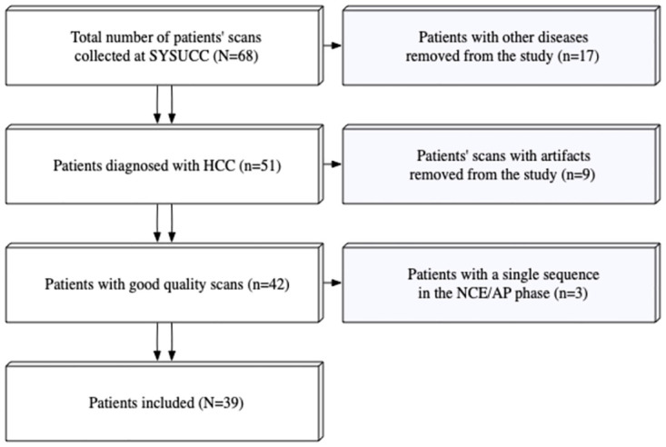
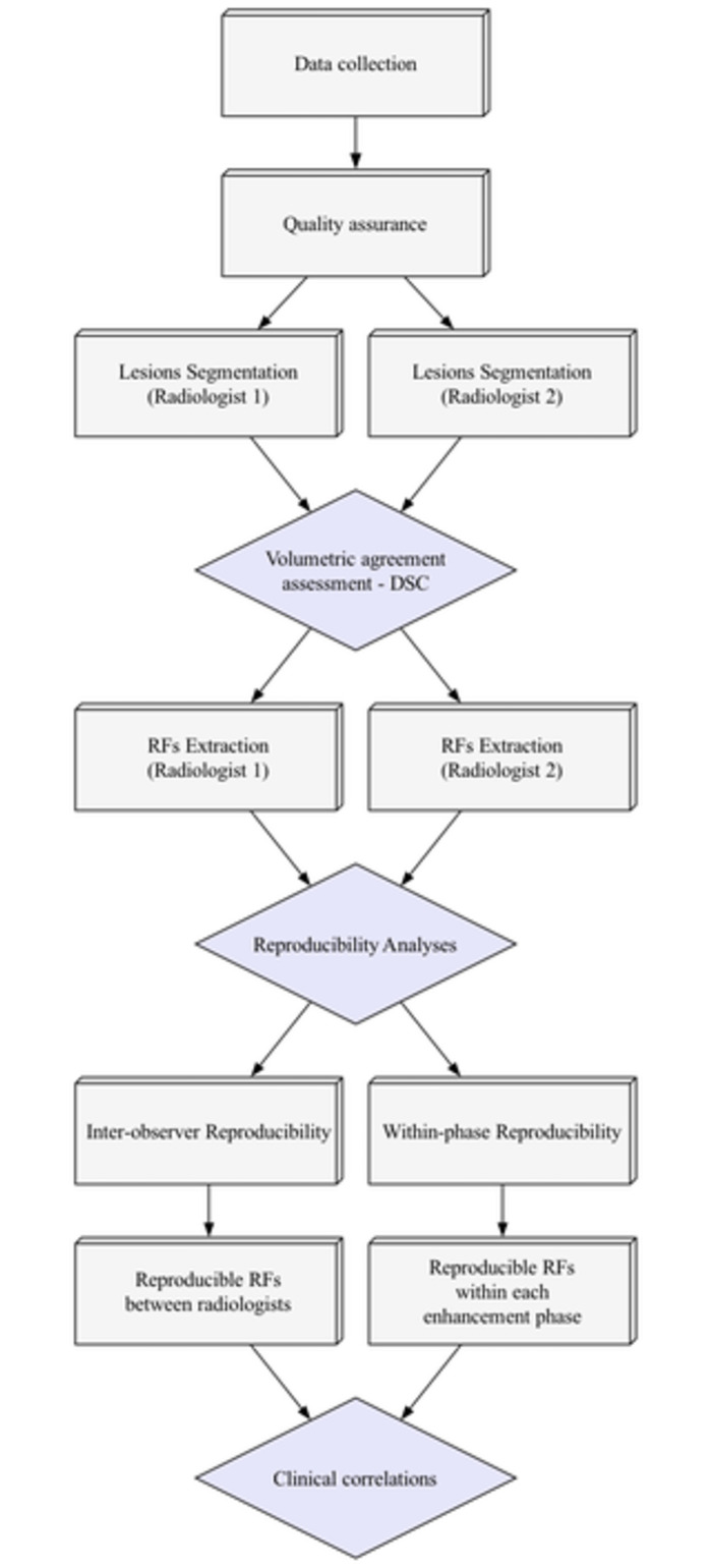
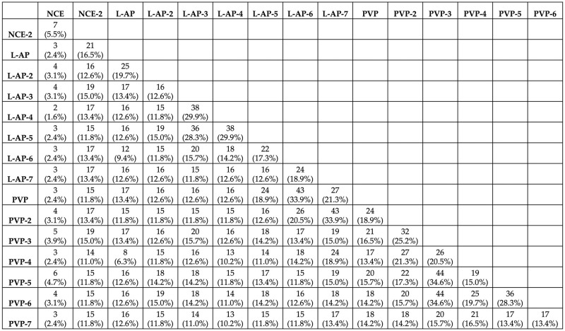
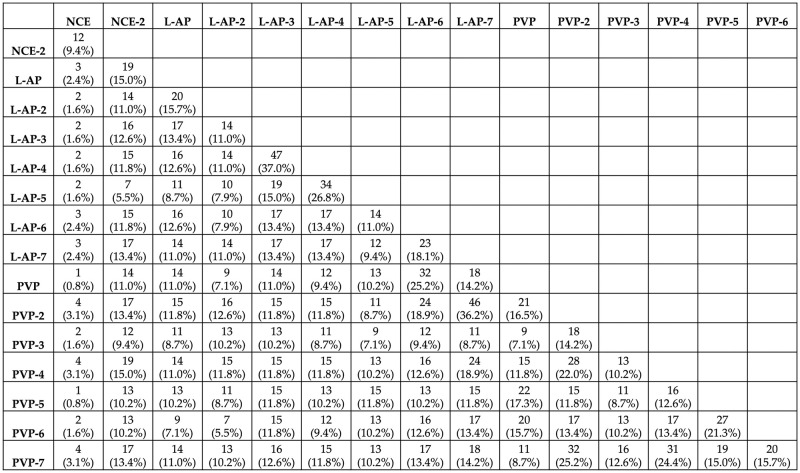
We analyzed a set of DCE-CT scans of 39 patients diagnosed with HCC. Two radiologists independently segmented the scans, and RFs were extracted from each sequence of the DCE-CT scans. The same lesion was segmented across the DCE-CT sequences of each patient's scan. From each lesion, 127 commonly used RFs were extracted. The reproducibility of RFs was assessed with regard to (i) inter-observer variability, by evaluating the reproducibility of RFs between the two radiologists; and (ii) timing of acquisition following contrast injection (inter- and intra-imaging phase). The reproducibility of RFs was assessed using the concordance correlation coefficient (CCC), with a cut-off value of 0.90. Reproducible RFs were used for building XGBoost classification models for the differentiation of HCC differentiation.
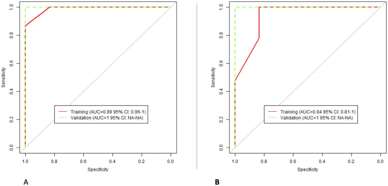
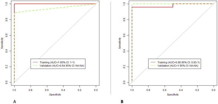
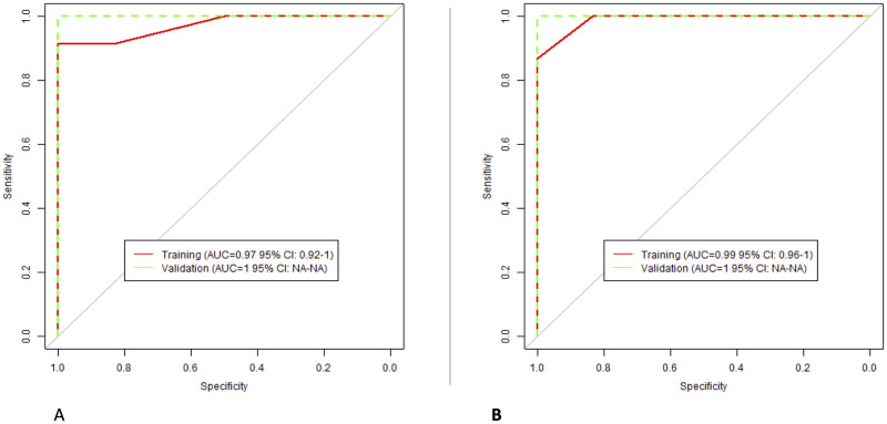
Inter-observer analyses across the different contrast-enhancement phases showed that the number of reproducible RFs was 29 (22.8%), 52 (40.9%), and 36 (28.3%) for the non-contrast enhanced, late arterial, and portal venous phases, respectively. Intra- and inter-sequence analyses revealed that the number of reproducible RFs ranged between 1 (0.8%) and 47 (37%), inversely related with time interval between the sequences. XGBoost algorithms built using reproducible RFs in each phase were found to be high predictive ability of the degree of HCC tumor differentiation.
The reproducibility of many RFs was significantly impacted by inter-observer variability, and a larger number of RFs were impacted by the difference in the time of acquisition after contrast injection. Our findings highlight the need for quality assessment to ensure that scans are analyzed in the same physiologic imaging phase in quantitative imaging studies, or that phase-wide reproducible RFs are selected. Overall, the study emphasizes the importance of reproducibility and quality control when using RFs as biomarkers for clinical applications.
评估从经动态对比增强 CT(DCE-CT)扫描诊断为肝细胞癌(HCC)的患者中提取的放射组学特征(RFs)的可重复性,具体涉及观察者间变异性和对比剂注射后采集时间。还研究了可重复 RFs 对区分 HCC 分化程度的预测能力。
我们分析了一组 39 例经 HCC 诊断的患者的 DCE-CT 扫描。两位放射科医生分别对扫描进行分割,并从每个 DCE-CT 序列中提取 RFs。对每位患者的扫描中 DCE-CT 序列中的相同病变进行分割。从每个病变中提取 127 个常用 RFs。通过评估两位放射科医生之间 RFs 的可重复性,评估 RFs 的可重复性(i)观察者间变异性;并通过评估对比剂注射后(不同成像期和同成像期)采集的时间评估 RFs 的可重复性。使用一致性相关系数(CCC)评估 RFs 的可重复性,截断值为 0.90。使用 XGBoost 分类模型对 HCC 分化进行区分,可重复性 RFs 用于构建该模型。
不同对比增强阶段的观察者间分析表明,非增强期、晚期动脉期和门静脉期的可重复性 RFs 数量分别为 29(22.8%)、52(40.9%)和 36(28.3%)。序列内和序列间分析表明,可重复性 RFs 的数量在 1(0.8%)和 47(37%)之间变化,与序列之间的时间间隔呈反比关系。使用每个阶段的可重复性 RFs 构建的 XGBoost 算法对 HCC 肿瘤分化程度具有较高的预测能力。
许多 RFs 的可重复性受到观察者间变异性的显著影响,更多的 RFs 受到对比剂注射后采集时间差异的影响。我们的发现强调了在定量成像研究中,为确保扫描在相同的生理成像期进行分析,或者选择全相可重复性 RFs,需要进行质量评估。总体而言,该研究强调了在临床应用中使用 RFs 作为生物标志物时,可重复性和质量控制的重要性。