Dehdab Reza, Brendlin Andreas S, Grözinger Gerd, Almansour Haidara, Brendel Jan Michael, Gassenmaier Sebastian, Ghibes Patrick, Werner Sebastian, Nikolaou Konstantin, Afat Saif
Department of Diagnostic and Interventional Radiology, University Hospital Tübingen, D-72076 Tuebingen, Germany.
Diagnostics (Basel). 2024 Sep 9;14(17):1989. doi: 10.3390/diagnostics14171989.
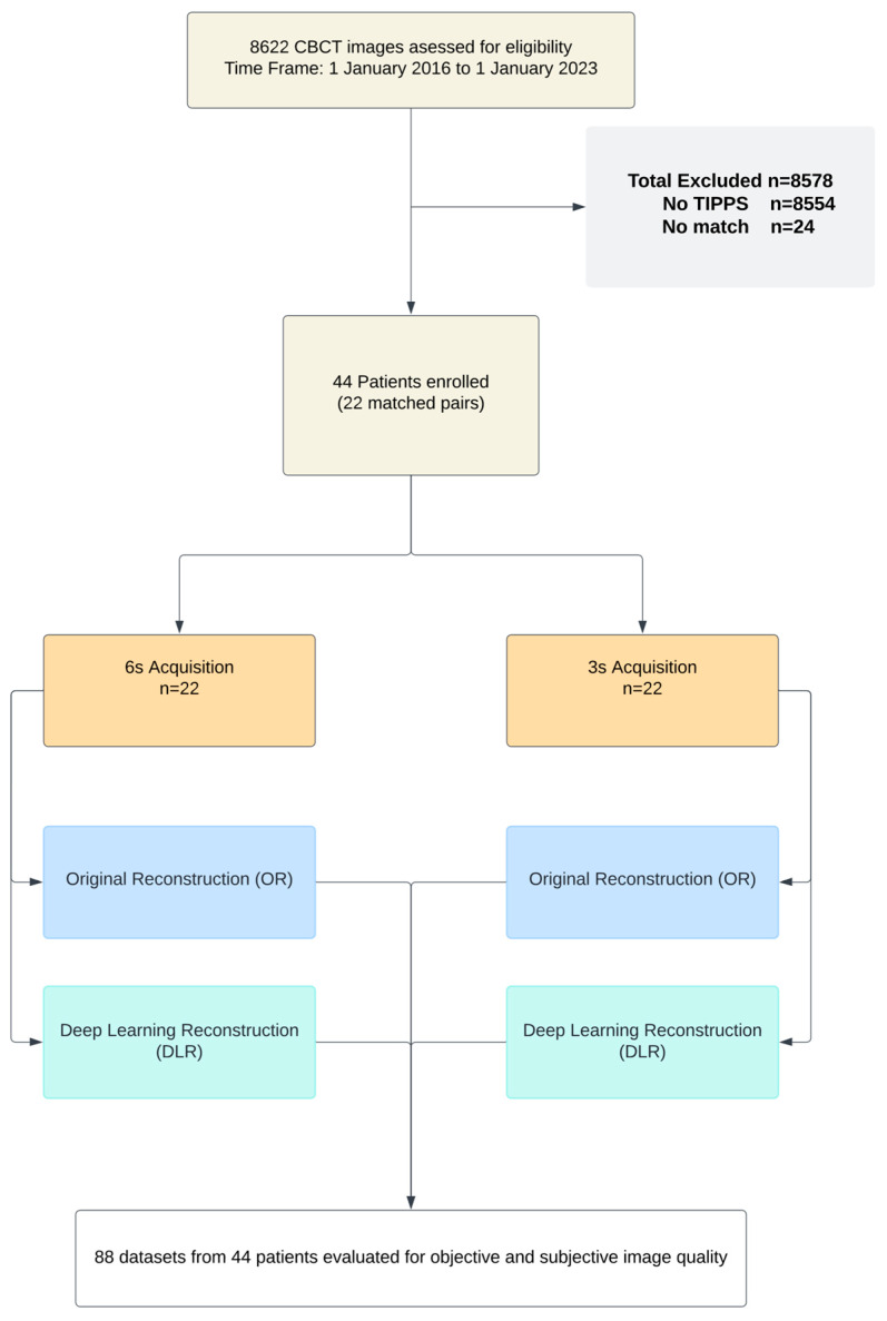
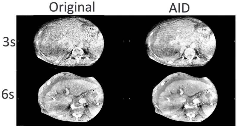
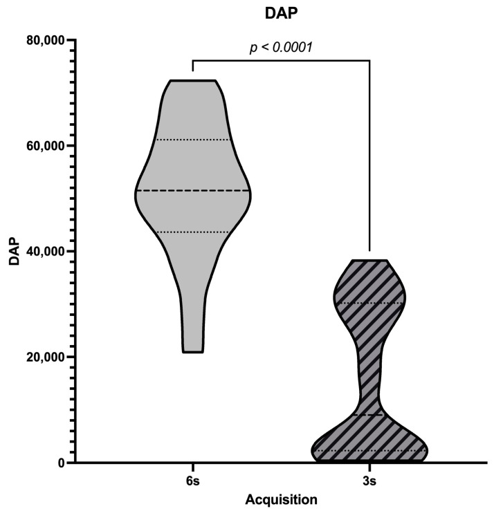
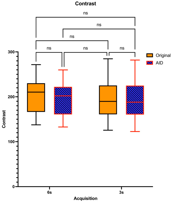
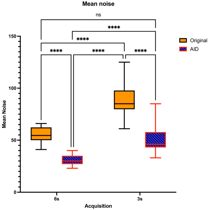
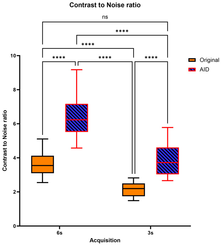
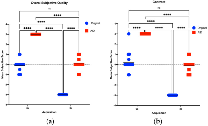
This study evaluates a deep learning-based denoising algorithm to improve the trade-off between radiation dose, image noise, and motion artifacts in TIPSS procedures, aiming for shorter acquisition times and reduced radiation with maintained diagnostic quality. In this retrospective study, TIPSS patients were divided based on CBCT acquisition times of 6 s and 3 s. Traditional weighted filtered back projection (Original) and an AI denoising algorithm (AID) were used for image reconstructions. Objective assessments of image quality included contrast, noise levels, and contrast-to-noise ratios (CNRs) through place-consistent region-of-interest (ROI) measurements across various critical areas pertinent to the TIPSS procedure. Subjective assessments were conducted by two blinded radiologists who evaluated the overall image quality, sharpness, contrast, and motion artifacts for each dataset combination. Statistical significance was determined using a mixed-effects model ( ≤ 0.05). From an initial cohort of 60 TIPSS patients, 44 were selected and paired. The mean dose-area product (DAP) for the 6 s acquisitions was 5138.50 ± 1325.57 µGy·m, significantly higher than the 2514.06 ± 691.59 µGym obtained for the 3 s series. CNR was highest in the 6 s-AID series ( < 0.05). Both denoised and original series showed consistent contrast for 6 s and 3 s acquisitions, with no significant noise differences between the 6 s Original and 3 s AID images ( > 0.9). Subjective assessments indicated superior quality in 6 s-AID images, with no significant overall quality difference between the 6 s-Original and 3 s-AID series ( > 0.9). The AI denoising algorithm enhances CBCT image quality in TIPSS procedures, allowing for shorter scans that reduce radiation exposure and minimize motion artifacts.
本研究评估了一种基于深度学习的去噪算法,以改善经颈静脉肝内门体分流术(TIPSS)中辐射剂量、图像噪声和运动伪影之间的权衡,目标是缩短采集时间并在保持诊断质量的同时减少辐射。在这项回顾性研究中,TIPSS患者根据6秒和3秒的CBCT采集时间进行分组。使用传统的加权滤波反投影(原始方法)和人工智能去噪算法(AID)进行图像重建。通过在与TIPSS手术相关的各个关键区域进行位置一致的感兴趣区域(ROI)测量,对图像质量进行客观评估,包括对比度、噪声水平和对比噪声比(CNR)。由两名不知情的放射科医生进行主观评估,他们对每个数据集组合的整体图像质量、清晰度、对比度和运动伪影进行评估。使用混合效应模型确定统计学显著性(≤0.05)。从最初的60名TIPSS患者队列中,选择了44名并进行配对。6秒采集的平均剂量面积乘积(DAP)为5138.50±1325.57µGy·m,显著高于3秒系列的2514.06±691.59µGy·m。CNR在6秒-AID系列中最高(<0.05)。去噪系列和原始系列在6秒和3秒采集中均显示出一致的对比度,6秒原始图像和3秒AID图像之间的噪声没有显著差异(>0.9)。主观评估表明6秒-AID图像质量更优,6秒-原始系列和3秒-AID系列之间的整体质量没有显著差异(>0.9)。人工智能去噪算法提高了TIPSS手术中CBCT图像的质量,允许进行更短的扫描,从而减少辐射暴露并将运动伪影降至最低。