Department of Gastroenterology and Hepatology, Okayama University Graduate School of Medicine, Dentistry and Pharmaceutical Science, 2-5-1, Shikata-Cho, Kita-Ku, Okayama, Okayama, Japan.
Department of Pathology, Okayama University Graduate School of Medicine, Dentistry and Pharmaceutical Science, Okayama, Japan.
Sci Rep. 2024 Sep 28;14(1):22441. doi: 10.1038/s41598-024-72312-3.
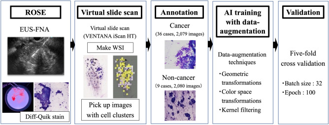
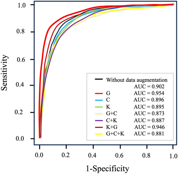
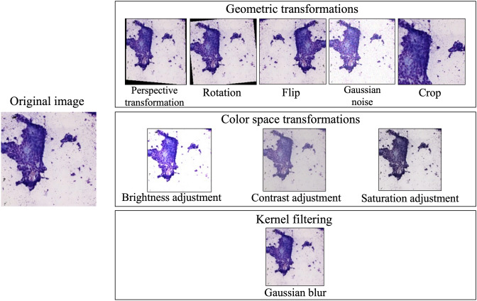
Rapid on-site cytopathology evaluation (ROSE) has been considered an effective method to increase the diagnostic ability of endoscopic ultrasound-guided fine needle aspiration (EUS-FNA); however, ROSE is unavailable in most institutes worldwide due to the shortage of cytopathologists. To overcome this situation, we created an artificial intelligence (AI)-based system (the ROSE-AI system), which was trained with the augmented data to evaluate the slide images acquired by EUS-FNA. This study aimed to clarify the effects of such data-augmentation on establishing an effective ROSE-AI system by comparing the efficacy of various data-augmentation techniques. The ROSE-AI system was trained with increased data obtained by the various data-augmentation techniques, including geometric transformation, color space transformation, and kernel filtering. By performing five-fold cross-validation, we compared the efficacy of each data-augmentation technique on the increasing diagnostic abilities of the ROSE-AI system. We collected 4059 divided EUS-FNA slide images from 36 patients with pancreatic cancer and nine patients with non-pancreatic cancer. The diagnostic ability of the ROSE-AI system without data augmentation had a sensitivity, specificity, and accuracy of 87.5%, 79.7%, and 83.7%, respectively. While, some data-augmentation techniques decreased diagnostic ability, the ROSE-AI system trained only with the augmented data using the geometric transformation technique had the highest diagnostic accuracy (88.2%). We successfully developed a prototype ROSE-AI system with high diagnostic ability. Each data-augmentation technique may have various compatibilities with AI-mediated diagnostics, and the geometric transformation was the most effective for the ROSE-AI system.
快速现场细胞学评估 (ROSE) 已被认为是提高内镜超声引导下细针抽吸 (EUS-FNA) 诊断能力的有效方法;然而,由于细胞病理学家短缺,世界上大多数机构都无法进行 ROSE。为了克服这种情况,我们创建了一个基于人工智能 (AI) 的系统(ROSE-AI 系统),该系统使用扩充数据来评估 EUS-FNA 获得的幻灯片图像。本研究旨在通过比较各种数据增强技术的效果,阐明数据增强对建立有效的 ROSE-AI 系统的影响。ROSE-AI 系统使用通过各种数据增强技术获得的扩充数据进行训练,包括几何变换、颜色空间变换和核滤波。通过进行五重交叉验证,我们比较了每种数据增强技术对提高 ROSE-AI 系统诊断能力的效果。我们从 36 名胰腺癌患者和 9 名非胰腺癌患者中收集了 4059 张分割的 EUS-FNA 幻灯片图像。未经数据增强的 ROSE-AI 系统的诊断能力具有 87.5%、79.7%和 83.7%的敏感性、特异性和准确性。虽然有些数据增强技术降低了诊断能力,但仅使用几何变换技术对增强数据进行训练的 ROSE-AI 系统具有最高的诊断准确性(88.2%)。我们成功开发了一种具有高诊断能力的原型 ROSE-AI 系统。每种数据增强技术可能与 AI 介导的诊断具有不同的兼容性,而几何变换对 ROSE-AI 系统最有效。