• 文献检索
  • 文档翻译
  • 深度研究
  • 学术资讯
  • Suppr Zotero 插件Zotero 插件
  • 邀请有礼
  • 套餐&价格
  • 历史记录
应用&插件
Suppr Zotero 插件Zotero 插件浏览器插件Mac 客户端Windows 客户端微信小程序
定价
高级版会员购买积分包购买API积分包
服务
文献检索文档翻译深度研究API 文档MCP 服务
关于我们
关于 Suppr公司介绍联系我们用户协议隐私条款
关注我们

Suppr 超能文献

核心技术专利:CN118964589B侵权必究
粤ICP备2023148730 号-1Suppr @ 2026

文献检索

告别复杂PubMed语法,用中文像聊天一样搜索,搜遍4000万医学文献。AI智能推荐,让科研检索更轻松。

立即免费搜索

文件翻译

保留排版,准确专业,支持PDF/Word/PPT等文件格式,支持 12+语言互译。

免费翻译文档

深度研究

AI帮你快速写综述,25分钟生成高质量综述,智能提取关键信息,辅助科研写作。

立即免费体验

通过低相干全层析成像对活的未标记的小肠类器官进行长期的三维高分辨率成像。

Long-term three-dimensional high-resolution imaging of live unlabeled small intestinal organoids via low-coherence holotomography.

机构信息

Graduate School of Medical Science and Engineering, Korea Advanced Institute of Science and Technology (KAIST), Daejeon, 34141, Republic of Korea.

KAIST Institute for Health Science and Technology, Daejeon, 34141, Republic of Korea.

出版信息

Exp Mol Med. 2024 Oct;56(10):2162-2170. doi: 10.1038/s12276-024-01312-0. Epub 2024 Oct 1.

DOI:10.1038/s12276-024-01312-0
PMID:39349827
原文链接:https://pmc.ncbi.nlm.nih.gov/articles/PMC11541879/
Abstract

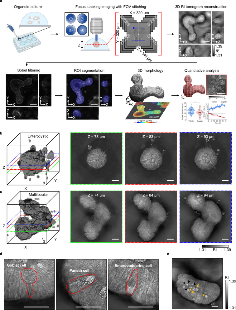
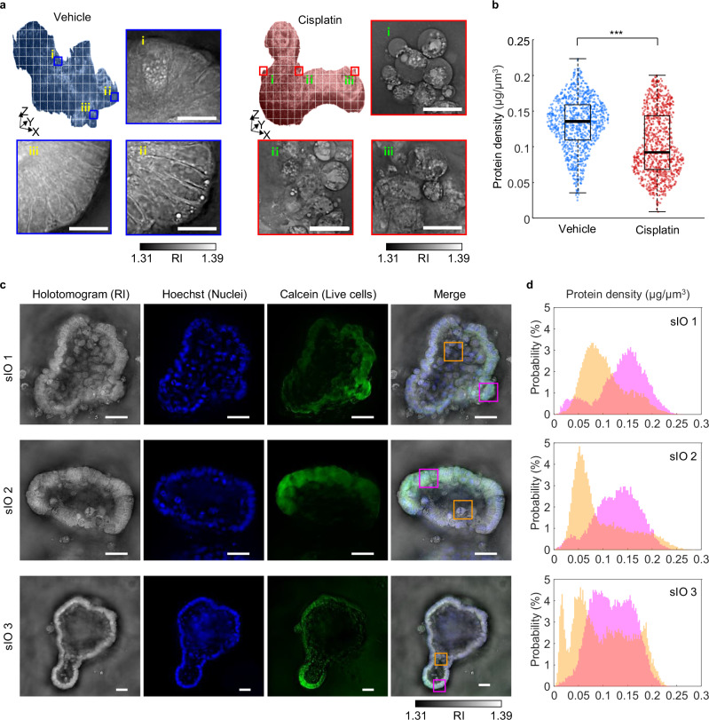
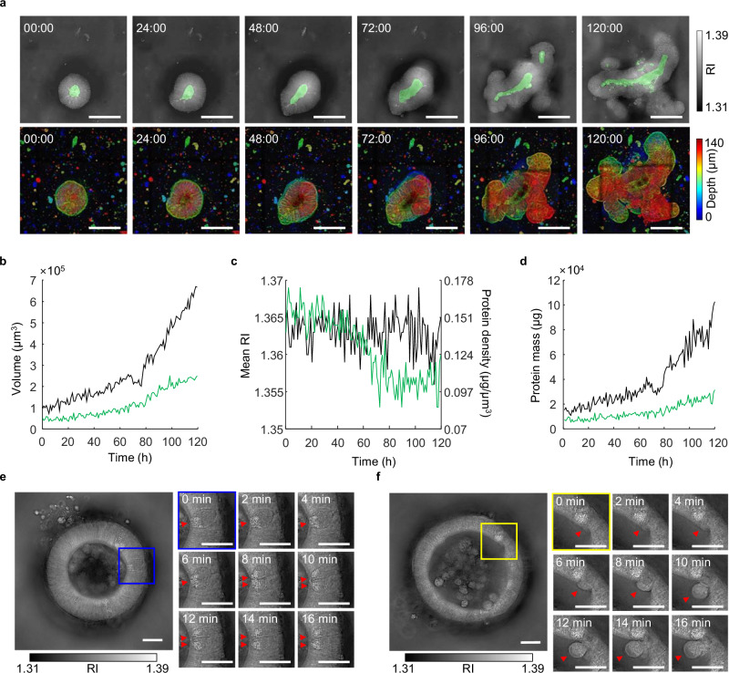
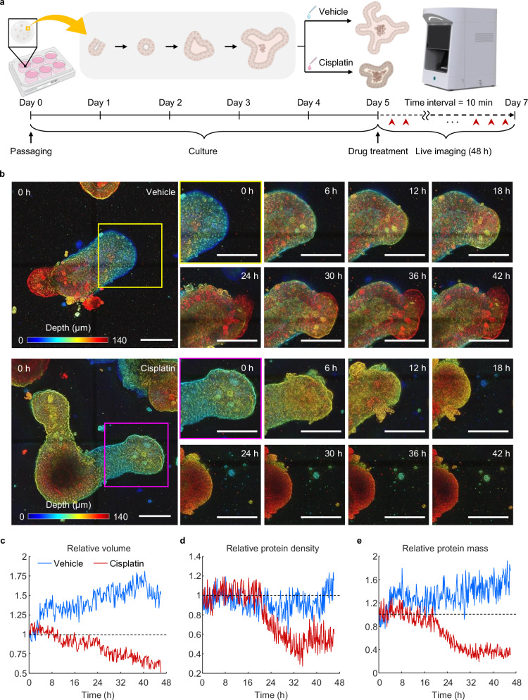
Organoids, which are miniature in vitro versions of organs, possess significant potential for studying human diseases and elucidating their underlying mechanisms. Live imaging techniques play a crucial role in organoid research and contribute to elucidating the complex structure and dynamic biological phenomena of organoids. However, live, unlabeled high-resolution imaging of native organoids is challenging, primarily owing to the complexities of sample handling and optical scattering inherent in three-dimensional (3D) structures. Additionally, conventional imaging methods fail to capture the real-time dynamic processes of growing organoids. In this study, we introduce low-coherence holotomography as an advanced, label-free, quantitative imaging modality designed to overcome several technical obstacles for long-term live imaging of 3D organoids. We demonstrate the efficacy of low-coherence holotomography by capturing high-resolution morphological details and dynamic activities within mouse small intestinal organoids at subcellular resolution. Moreover, our approach facilitates the distinction between viable and nonviable organoids, significantly enhancing its utility in organoid-based research. This advancement underscores the critical role of live imaging in organoid studies, offering a more comprehensive understanding of these complex systems.

摘要

类器官是微型的体外器官模型,在研究人类疾病和阐明其潜在机制方面具有重要潜力。活细胞成像技术在类器官研究中起着至关重要的作用,有助于阐明类器官的复杂结构和动态生物学现象。然而,对天然类器官进行活细胞、无标记的高分辨率成像具有挑战性,主要是由于三维(3D)结构中样品处理和光散射的复杂性。此外,传统的成像方法无法捕捉到正在生长的类器官的实时动态过程。在本研究中,我们引入低相干全层析成像作为一种先进的、无标记的定量成像方式,旨在克服长期对 3D 类器官进行活细胞成像的若干技术障碍。我们通过在亚细胞分辨率下捕获小鼠小肠类器官的高分辨率形态细节和动态活动,证明了低相干全层析成像的有效性。此外,我们的方法能够区分有活力和无活力的类器官,极大地提高了其在基于类器官的研究中的应用价值。这一进展突显了活细胞成像在类器官研究中的关键作用,为深入了解这些复杂系统提供了更全面的认识。

https://cdn.ncbi.nlm.nih.gov/pmc/blobs/1f35/11541879/3571daef7eb9/12276_2024_1312_Fig4_HTML.jpg
https://cdn.ncbi.nlm.nih.gov/pmc/blobs/1f35/11541879/611acf38f8dc/12276_2024_1312_Fig1_HTML.jpg
https://cdn.ncbi.nlm.nih.gov/pmc/blobs/1f35/11541879/238b189dd191/12276_2024_1312_Fig2_HTML.jpg
https://cdn.ncbi.nlm.nih.gov/pmc/blobs/1f35/11541879/34c26b6dd49d/12276_2024_1312_Fig3_HTML.jpg
https://cdn.ncbi.nlm.nih.gov/pmc/blobs/1f35/11541879/3571daef7eb9/12276_2024_1312_Fig4_HTML.jpg

相似文献

1
Long-term three-dimensional high-resolution imaging of live unlabeled small intestinal organoids via low-coherence holotomography.通过低相干全层析成像对活的未标记的小肠类器官进行长期的三维高分辨率成像。
Exp Mol Med. 2024 Oct;56(10):2162-2170. doi: 10.1038/s12276-024-01312-0. Epub 2024 Oct 1.
2
Non-invasive label-free imaging analysis pipeline for in situ characterization of 3D brain organoids.用于 3D 脑类器官原位特征分析的无创无标记成像分析流程。
Sci Rep. 2024 Sep 27;14(1):22331. doi: 10.1038/s41598-024-72038-2.
3
Long-term culture-induced phenotypic difference and efficient cryopreservation of small intestinal organoids by treatment timing of Rho kinase inhibitor.通过Rho激酶抑制剂处理时间实现小肠类器官的长期培养诱导表型差异及高效冷冻保存
World J Gastroenterol. 2017 Feb 14;23(6):964-975. doi: 10.3748/wjg.v23.i6.964.
4
A fully automated high-throughput workflow for 3D-based chemical screening in human midbrain organoids.一种全自动高通量工作流程,用于基于 3D 的人类中脑细胞类器官中的化学筛选。
Elife. 2020 Nov 3;9:e52904. doi: 10.7554/eLife.52904.
5
Live-Cell Calcium Imaging in 3D Intestinal Organoids.3D 肠道类器官中的活细胞钙成像。
Methods Mol Biol. 2025;2861:213-221. doi: 10.1007/978-1-0716-4164-4_16.
6
High-Resolution Imaging of Spindle Orientation Dynamics in 3D Intestinal Organoids.高分辨率成像三维肠道类器官中纺锤体取向动力学。
Methods Mol Biol. 2025;2872:257-267. doi: 10.1007/978-1-0716-4224-5_18.
7
Single-Cell Resolution Three-Dimensional Imaging of Intact Organoids.完整类器官的单细胞分辨率三维成像
J Vis Exp. 2020 Jun 5(160). doi: 10.3791/60709.
8
Deep learning based characterization of human organoids using optical coherence tomography.基于深度学习的光学相干断层扫描对人类类器官的表征
Biomed Opt Express. 2024 Apr 17;15(5):3112-3127. doi: 10.1364/BOE.515781. eCollection 2024 May 1.
9
Establishment and Culture of Human Intestinal Organoids Derived from Adult Stem Cells.人肠类器官的建立和培养来源于成体干细胞。
Curr Protoc Immunol. 2020 Sep;130(1):e106. doi: 10.1002/cpim.106.
10
[Optimization of the culture system for chicken small intestinal organoids].[鸡小肠类器官培养系统的优化]
Sheng Wu Gong Cheng Xue Bao. 2024 Dec 25;40(12):4645-4659. doi: 10.13345/j.cjb.240331.

引用本文的文献

1
Gastrointestinal tract, its pathophysiology and models: A "quick" reference guide to translational studies.胃肠道、其病理生理学及模型:转化研究的“快速”参考指南。
World J Gastroenterol. 2025 Jul 28;31(28):108297. doi: 10.3748/wjg.v31.i28.108297.
2
Precision medicine research progress based on colorectal cancer organoids.基于结直肠癌类器官的精准医学研究进展
Discov Oncol. 2025 Jun 23;16(1):1181. doi: 10.1007/s12672-025-02959-5.
3
The midbody and midbody remnant: from cellular debris to signaling organelle with diagnostic and therapeutic potential.

本文引用的文献

1
Perspective on quantitative phase imaging to improve precision cancer medicine.定量相位成像在提高精准癌症医学中的应用展望。
J Biomed Opt. 2024 Jun;29(Suppl 2):S22705. doi: 10.1117/1.JBO.29.S2.S22705. Epub 2024 Mar 26.
2
Artificial intelligence-enabled quantitative phase imaging methods for life sciences.人工智能赋能的生命科学定量相位成像方法。
Nat Methods. 2023 Nov;20(11):1645-1660. doi: 10.1038/s41592-023-02041-4. Epub 2023 Oct 23.
3
In-silico clearing approach for deep refractive index tomography by partial reconstruction and wave-backpropagation.
中间体及中间体残余物:从细胞碎片到具有诊断和治疗潜力的信号细胞器
Mol Biol Cell. 2025 Jul 1;36(7):re4. doi: 10.1091/mbc.E25-03-0120. Epub 2025 May 28.
4
Advances and Challenges in Modeling Autosomal Dominant Polycystic Kidney Disease: A Focus on Kidney Organoids.常染色体显性多囊肾病建模的进展与挑战:聚焦肾类器官
Biomedicines. 2025 Feb 19;13(2):523. doi: 10.3390/biomedicines13020523.
5
Navigating the Complexity of Resistance in Lung Cancer Therapy: Mechanisms, Organoid Models, and Strategies for Overcoming Treatment Failure.应对肺癌治疗中耐药性的复杂性:机制、类器官模型及克服治疗失败的策略
Cancers (Basel). 2024 Nov 28;16(23):3996. doi: 10.3390/cancers16233996.
6
Novel imaging and biophysical approaches to study tissue hydraulics in mammalian folliculogenesis.用于研究哺乳动物卵泡发生过程中组织水力学的新型成像和生物物理方法。
Biophys Rev. 2024 Oct 7;16(5):625-637. doi: 10.1007/s12551-024-01231-4. eCollection 2024 Oct.
7
Perspective on quantitative phase imaging to improve precision cancer medicine.定量相位成像在提高精准癌症医学中的应用展望。
J Biomed Opt. 2024 Jun;29(Suppl 2):S22705. doi: 10.1117/1.JBO.29.S2.S22705. Epub 2024 Mar 26.
基于部分重建和波反向传播的深度折射率层析成像的计算机模拟清除方法。
Light Sci Appl. 2023 Apr 27;12(1):101. doi: 10.1038/s41377-023-01144-z.
4
Deep learning-enabled virtual histological staining of biological samples.基于深度学习的生物样本虚拟组织学染色
Light Sci Appl. 2023 Mar 3;12(1):57. doi: 10.1038/s41377-023-01104-7.
5
Stain-free identification of cell nuclei using tomographic phase microscopy in flow cytometry.在流式细胞术中使用断层相显微镜对细胞核进行无标记识别。
Nat Photonics. 2022 Dec;16(12):851-859. doi: 10.1038/s41566-022-01096-7. Epub 2022 Nov 10.
6
Multiscale light-sheet organoid imaging framework.多尺度光片器官成像框架。
Nat Commun. 2022 Aug 18;13(1):4864. doi: 10.1038/s41467-022-32465-z.
7
Organoid Imaging: Seeing Development and Function.类器官成像:观察发育与功能
Annu Rev Cell Dev Biol. 2022 Oct 6;38:447-466. doi: 10.1146/annurev-cellbio-120320-035146. Epub 2022 Jun 29.
8
Present Application and Perspectives of Organoid Imaging Technology.类器官成像技术的当前应用与前景
Bioengineering (Basel). 2022 Mar 16;9(3):121. doi: 10.3390/bioengineering9030121.
9
Label-free multiplexed microtomography of endogenous subcellular dynamics using generalizable deep learning.基于可推广深度学习的无标记多路复用微层析术用于内源性亚细胞动力学研究。
Nat Cell Biol. 2021 Dec;23(12):1329-1337. doi: 10.1038/s41556-021-00802-x. Epub 2021 Dec 7.
10
Optimizing illumination in three-dimensional deconvolution microscopy for accurate refractive index tomography.优化三维反卷积显微镜的照明以实现精确的折射率层析成像。
Opt Express. 2021 Mar 1;29(5):6293-6301. doi: 10.1364/OE.412510.