• 文献检索
  • 文档翻译
  • 深度研究
  • 学术资讯
  • Suppr Zotero 插件Zotero 插件
  • 邀请有礼
  • 套餐&价格
  • 历史记录
应用&插件
Suppr Zotero 插件Zotero 插件浏览器插件Mac 客户端Windows 客户端微信小程序
定价
高级版会员购买积分包购买API积分包
服务
文献检索文档翻译深度研究API 文档MCP 服务
关于我们
关于 Suppr公司介绍联系我们用户协议隐私条款
关注我们

Suppr 超能文献

核心技术专利:CN118964589B侵权必究
粤ICP备2023148730 号-1Suppr @ 2026

文献检索

告别复杂PubMed语法,用中文像聊天一样搜索,搜遍4000万医学文献。AI智能推荐,让科研检索更轻松。

立即免费搜索

文件翻译

保留排版,准确专业,支持PDF/Word/PPT等文件格式,支持 12+语言互译。

免费翻译文档

深度研究

AI帮你快速写综述,25分钟生成高质量综述,智能提取关键信息,辅助科研写作。

立即免费体验

基于空间注意力的 CSR-Unet 框架,用于使用 CT 图像进行硬膜下和硬膜外血肿分割和分类。

Spatial attention-based CSR-Unet framework for subdural and epidural hemorrhage segmentation and classification using CT images.

机构信息

School of Electronics Engineering, Vellore Institute of Technology, Vellore, India.

出版信息

BMC Med Imaging. 2024 Oct 22;24(1):285. doi: 10.1186/s12880-024-01455-6.

DOI:10.1186/s12880-024-01455-6
PMID:39438833
原文链接:https://pmc.ncbi.nlm.nih.gov/articles/PMC11494839/
Abstract

BACKGROUND

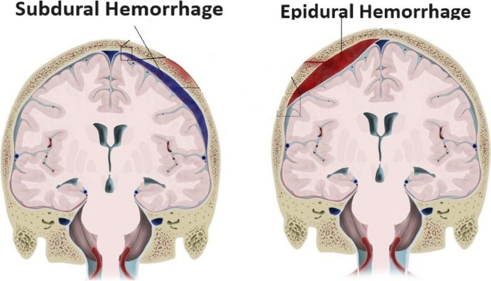
Automatic diagnosis and brain hemorrhage segmentation in Computed Tomography (CT) may be helpful in assisting the neurosurgeon in developing treatment plans that improve the patient's chances of survival. Because medical segmentation of images is important and performing operations manually is challenging, many automated algorithms have been developed for this purpose, primarily focusing on certain image modalities. Whenever a blood vessel bursts, a dangerous medical condition known as intracranial hemorrhage (ICH) occurs. For best results, quick action is required. That being said, identifying subdural (SDH) and epidural haemorrhages (EDH) is a difficult task in this field and calls for a new, more precise detection method.

METHODS

This work uses a head CT scan to detect cerebral bleeding and distinguish between two types of dural hemorrhages using deep learning techniques. This paper proposes a rich segmentation approach to segment both SDH and EDH by enhancing segmentation efficiency with a better feature extraction procedure. This method incorporates Spatial attention- based CSR (convolution-SE-residual) Unet, for rich segmentation and precise feature extraction.

RESULTS

According to the study's findings, the CSR based Spatial network performs better than the other models, exhibiting impressive metrics for all assessed parameters with a mean dice coefficient of 0.970 and mean IoU of 0.718, while EDH and SDH dice scores are 0.983 and 0.969 respectively.

CONCLUSIONS

The CSR Spatial network experiment results show that it can perform well regarding dice coefficient. Furthermore, Spatial Unet based on CSR may effectively model the complicated in segmentations and rich feature extraction and improve the representation learning compared to alternative deep learning techniques, of illness and medical treatment, to enhance the meticulousness in predicting the fatality.

摘要

背景

在计算机断层扫描(CT)中自动诊断和脑出血分割可能有助于神经外科医生制定改善患者生存机会的治疗计划。由于医学图像分割很重要,手动操作具有挑战性,因此已经开发了许多自动化算法来实现这一目的,主要集中在某些图像模式上。每当血管破裂时,就会发生一种称为颅内出血(ICH)的危险医疗状况。为了获得最佳效果,需要迅速采取行动。也就是说,在该领域,识别硬膜下(SDH)和硬膜外血肿(EDH)是一项艰巨的任务,需要一种新的、更精确的检测方法。

方法

这项工作使用头部 CT 扫描来检测脑内出血,并使用深度学习技术区分两种硬膜下出血。本文提出了一种丰富的分割方法,通过使用更好的特征提取过程增强分割效率,对 SDH 和 EDH 进行分割。该方法结合了基于空间注意力的 CSR(卷积-SE-残差)Unet,用于丰富分割和精确特征提取。

结果

根据研究结果,基于 CSR 的空间网络的表现优于其他模型,在所有评估参数上均表现出出色的指标,平均骰子系数为 0.970,平均 IoU 为 0.718,而 EDH 和 SDH 的骰子分数分别为 0.983 和 0.969。

结论

CSR 空间网络实验结果表明,它在骰子系数方面表现良好。此外,基于 CSR 的空间 U-Net 可以有效地对复杂的分割和丰富的特征提取进行建模,并通过与替代深度学习技术相比,提高表示学习能力,从而提高对疾病和治疗的预测准确性。

https://cdn.ncbi.nlm.nih.gov/pmc/blobs/6f35/11494839/846acdc28776/12880_2024_1455_Fig15_HTML.jpg
https://cdn.ncbi.nlm.nih.gov/pmc/blobs/6f35/11494839/4a1f4a8b0a9f/12880_2024_1455_Fig1_HTML.jpg
https://cdn.ncbi.nlm.nih.gov/pmc/blobs/6f35/11494839/a4a6bab2b3bf/12880_2024_1455_Fig2_HTML.jpg
https://cdn.ncbi.nlm.nih.gov/pmc/blobs/6f35/11494839/bc929024bad4/12880_2024_1455_Fig3_HTML.jpg
https://cdn.ncbi.nlm.nih.gov/pmc/blobs/6f35/11494839/b800b957c54d/12880_2024_1455_Fig4_HTML.jpg
https://cdn.ncbi.nlm.nih.gov/pmc/blobs/6f35/11494839/dbbe571e2295/12880_2024_1455_Fig5_HTML.jpg
https://cdn.ncbi.nlm.nih.gov/pmc/blobs/6f35/11494839/2a5d29591871/12880_2024_1455_Fig6_HTML.jpg
https://cdn.ncbi.nlm.nih.gov/pmc/blobs/6f35/11494839/25fbb79b3874/12880_2024_1455_Fig7_HTML.jpg
https://cdn.ncbi.nlm.nih.gov/pmc/blobs/6f35/11494839/ecd009c98d0a/12880_2024_1455_Fig8_HTML.jpg
https://cdn.ncbi.nlm.nih.gov/pmc/blobs/6f35/11494839/5b27b8b7afa4/12880_2024_1455_Figa_HTML.jpg
https://cdn.ncbi.nlm.nih.gov/pmc/blobs/6f35/11494839/802122a37461/12880_2024_1455_Fig9_HTML.jpg
https://cdn.ncbi.nlm.nih.gov/pmc/blobs/6f35/11494839/76e5f4226ea1/12880_2024_1455_Fig10_HTML.jpg
https://cdn.ncbi.nlm.nih.gov/pmc/blobs/6f35/11494839/054a15ec6563/12880_2024_1455_Fig11_HTML.jpg
https://cdn.ncbi.nlm.nih.gov/pmc/blobs/6f35/11494839/f54a15278f9d/12880_2024_1455_Fig12_HTML.jpg
https://cdn.ncbi.nlm.nih.gov/pmc/blobs/6f35/11494839/2b022074b2fb/12880_2024_1455_Fig13_HTML.jpg
https://cdn.ncbi.nlm.nih.gov/pmc/blobs/6f35/11494839/6fd26786da3f/12880_2024_1455_Fig14_HTML.jpg
https://cdn.ncbi.nlm.nih.gov/pmc/blobs/6f35/11494839/846acdc28776/12880_2024_1455_Fig15_HTML.jpg

相似文献

1
Spatial attention-based CSR-Unet framework for subdural and epidural hemorrhage segmentation and classification using CT images.基于空间注意力的 CSR-Unet 框架,用于使用 CT 图像进行硬膜下和硬膜外血肿分割和分类。
BMC Med Imaging. 2024 Oct 22;24(1):285. doi: 10.1186/s12880-024-01455-6.
2
A Robust Deep Learning Segmentation Method for Hematoma Volumetric Detection in Intracerebral Hemorrhage.一种用于脑出血血肿容量检测的稳健深度学习分割方法。
Stroke. 2022 Jan;53(1):167-176. doi: 10.1161/STROKEAHA.120.032243. Epub 2021 Oct 4.
3
Automatic hemorrhage segmentation on head CT scan for traumatic brain injury using 3D deep learning model.利用 3D 深度学习模型对头 CT 扫描外伤性脑损伤进行自动出血分割。
Comput Biol Med. 2022 Jul;146:105530. doi: 10.1016/j.compbiomed.2022.105530. Epub 2022 Apr 18.
4
Deep Network for the Automatic Segmentation and Quantification of Intracranial Hemorrhage on CT.用于CT图像上颅内出血自动分割与量化的深度网络
Front Neurosci. 2021 Jan 11;14:541817. doi: 10.3389/fnins.2020.541817. eCollection 2020.
5
ASD-Net: a novel U-Net based asymmetric spatial-channel convolution network for precise kidney and kidney tumor image segmentation.ASD-Net:一种新颖的基于 U-Net 的非对称空间-通道卷积网络,用于精确的肾脏和肾肿瘤图像分割。
Med Biol Eng Comput. 2024 Jun;62(6):1673-1687. doi: 10.1007/s11517-024-03025-y. Epub 2024 Feb 8.
6
Contralateral acute epidural hematoma after decompressive surgery of acute subdural hematoma: clinical features and outcome.急性硬膜下血肿减压术后对侧急性硬膜外血肿:临床特征与转归
J Trauma. 2008 Dec;65(6):1298-302. doi: 10.1097/TA.0b013e31815885d9.
7
A symmetric prior knowledge based deep learning model for intracerebral hemorrhage lesion segmentation.一种基于对称先验知识的脑出血病灶分割深度学习模型。
Front Physiol. 2022 Nov 23;13:977427. doi: 10.3389/fphys.2022.977427. eCollection 2022.
8
A Deep Learning-Based Automatic Segmentation and 3D Visualization Technique for Intracranial Hemorrhage Detection Using Computed Tomography Images.一种基于深度学习的自动分割与三维可视化技术,用于利用计算机断层扫描图像检测颅内出血。
Diagnostics (Basel). 2023 Jul 31;13(15):2537. doi: 10.3390/diagnostics13152537.
9
Eres-UNet++: Liver CT image segmentation based on high-efficiency channel attention and Res-UNet+.Eres-UNet++:基于高效通道注意力和Res-UNet+的肝脏CT图像分割
Comput Biol Med. 2023 May;158:106501. doi: 10.1016/j.compbiomed.2022.106501. Epub 2023 Jan 10.
10
Medical lesion segmentation by combining multimodal images with modality weighted UNet.基于模态加权 UNet 融合多模态图像的医学病灶分割。
Med Phys. 2022 Jun;49(6):3692-3704. doi: 10.1002/mp.15610. Epub 2022 Apr 7.

引用本文的文献

1
The association between hemoglobin-to-red blood cell distribution width ratio and 28-day mortality in epidural hemorrhage: a cohort study.血红蛋白与红细胞分布宽度比值与硬膜外出血患者28天死亡率的关系:一项队列研究
Front Neurol. 2025 Jul 8;16:1534098. doi: 10.3389/fneur.2025.1534098. eCollection 2025.
2
Intracranial hemorrhage segmentation and classification framework in computer tomography images using deep learning techniques.利用深度学习技术的计算机断层扫描图像中的颅内出血分割与分类框架
Sci Rep. 2025 May 17;15(1):17151. doi: 10.1038/s41598-025-01317-3.

本文引用的文献

1
Enhancing ECG-based heart age: impact of acquisition parameters and generalization strategies for varying signal morphologies and corruptions.增强基于心电图的心脏年龄:采集参数的影响以及针对不同信号形态和失真的泛化策略。
Front Cardiovasc Med. 2024 Jul 4;11:1424585. doi: 10.3389/fcvm.2024.1424585. eCollection 2024.
2
A deep convolutional neural network for the automatic segmentation of glioblastoma brain tumor: Joint spatial pyramid module and attention mechanism network.用于胶质母细胞瘤脑肿瘤自动分割的深度卷积神经网络:联合空间金字塔模块和注意力机制网络。
Artif Intell Med. 2024 Feb;148:102776. doi: 10.1016/j.artmed.2024.102776. Epub 2024 Jan 19.
3
Endoscopic Image Enhancement: Wavelet Transform and Guided Filter Decomposition-Based Fusion Approach.
内镜图像增强:基于小波变换和引导滤波分解的融合方法。
J Imaging. 2024 Jan 20;10(1):28. doi: 10.3390/jimaging10010028.
4
Unveiling the future of breast cancer assessment: a critical review on generative adversarial networks in elastography ultrasound.揭示乳腺癌评估的未来:弹性成像超声中生成对抗网络的批判性综述
Front Oncol. 2023 Dec 6;13:1282536. doi: 10.3389/fonc.2023.1282536. eCollection 2023.
5
Trends and patterns in the global burden of intracerebral hemorrhage: a comprehensive analysis from 1990 to 2019.全球脑出血负担的趋势与模式:1990年至2019年的综合分析
Front Neurol. 2023 Nov 21;14:1241158. doi: 10.3389/fneur.2023.1241158. eCollection 2023.
6
Estimating age and gender from electrocardiogram signals: A comprehensive review of the past decade.从心电图信号估计年龄和性别:过去十年的综合回顾。
Artif Intell Med. 2023 Dec;146:102690. doi: 10.1016/j.artmed.2023.102690. Epub 2023 Oct 21.
7
Automated Computer-Aided Detection and Classification of Intracranial Hemorrhage Using Ensemble Deep Learning Techniques.使用集成深度学习技术的颅内出血自动计算机辅助检测与分类
Diagnostics (Basel). 2023 Sep 18;13(18):2987. doi: 10.3390/diagnostics13182987.
8
A Coarse-to-Fine Fusion Network for Small Liver Tumor Detection and Segmentation: A Real-World Study.用于小肝脏肿瘤检测与分割的粗细融合网络:一项真实世界研究
Diagnostics (Basel). 2023 Jul 27;13(15):2504. doi: 10.3390/diagnostics13152504.
9
A systematic review on intracranial aneurysm and hemorrhage detection using machine learning and deep learning techniques.基于机器学习和深度学习技术的颅内动脉瘤与出血检测的系统评价。
Prog Biophys Mol Biol. 2023 Oct;183:1-16. doi: 10.1016/j.pbiomolbio.2023.07.001. Epub 2023 Jul 25.
10
A comparison of performance between a deep learning model with residents for localization and classification of intracranial hemorrhage.深度学习模型与住院医师在颅内出血定位和分类中的表现比较。
Sci Rep. 2023 Jun 20;13(1):9975. doi: 10.1038/s41598-023-37114-z.