• 文献检索
  • 文档翻译
  • 深度研究
  • 学术资讯
  • Suppr Zotero 插件Zotero 插件
  • 邀请有礼
  • 套餐&价格
  • 历史记录
应用&插件
Suppr Zotero 插件Zotero 插件浏览器插件Mac 客户端Windows 客户端微信小程序
定价
高级版会员购买积分包购买API积分包
服务
文献检索文档翻译深度研究API 文档MCP 服务
关于我们
关于 Suppr公司介绍联系我们用户协议隐私条款
关注我们

Suppr 超能文献

核心技术专利:CN118964589B侵权必究
粤ICP备2023148730 号-1Suppr @ 2026

文献检索

告别复杂PubMed语法,用中文像聊天一样搜索,搜遍4000万医学文献。AI智能推荐,让科研检索更轻松。

立即免费搜索

文件翻译

保留排版,准确专业,支持PDF/Word/PPT等文件格式,支持 12+语言互译。

免费翻译文档

深度研究

AI帮你快速写综述,25分钟生成高质量综述,智能提取关键信息,辅助科研写作。

立即免费体验

乳腺癌腋窝淋巴结转移风险列线图的构建与验证

Development and Validation of a Nomogram for Axillary Lymph Node Metastasis Risk in Breast Cancer.

作者信息

Wang Shijing, Zhang He, Wang Xin, Yu Juanhan, Zhang Qingfu, Zheng Yiwen, Zhang Tangbo, Mao Xiaoyun

机构信息

Department of Breast Surgery, The First Affiliated Hospital of China Medical University, Shenyang, Liaoning Province, 110001, China.

Department of Medical Imaging, Affiliated Hospital of Nantong University, Nantong, Jiangsu Province, 226001, China.

出版信息

J Cancer. 2024 Oct 7;15(18):6122-6134. doi: 10.7150/jca.100651. eCollection 2024.

DOI:10.7150/jca.100651
PMID:39440057
原文链接:https://pmc.ncbi.nlm.nih.gov/articles/PMC11493017/
Abstract

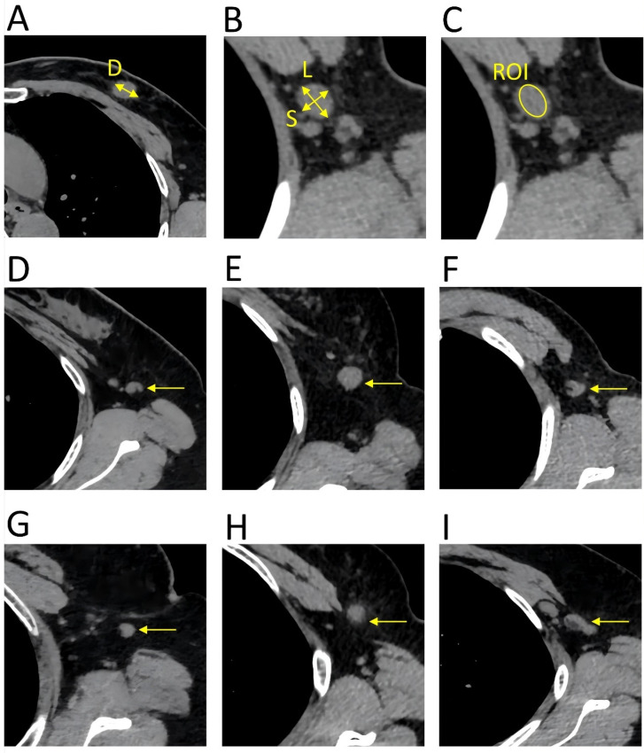
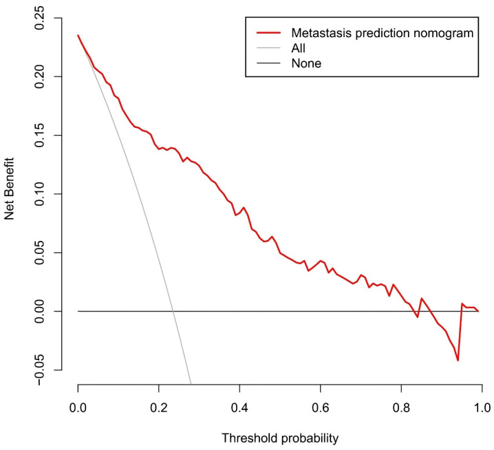
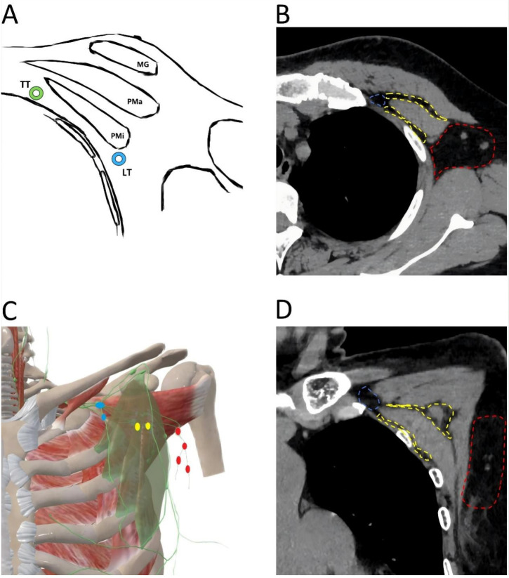
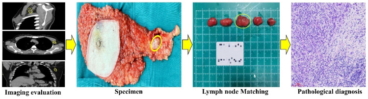
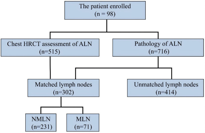
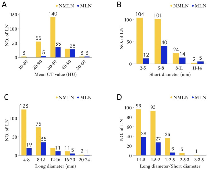
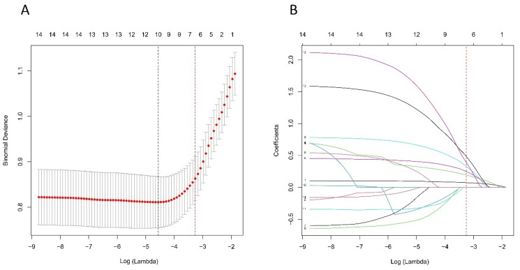
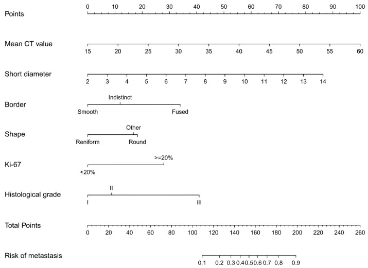
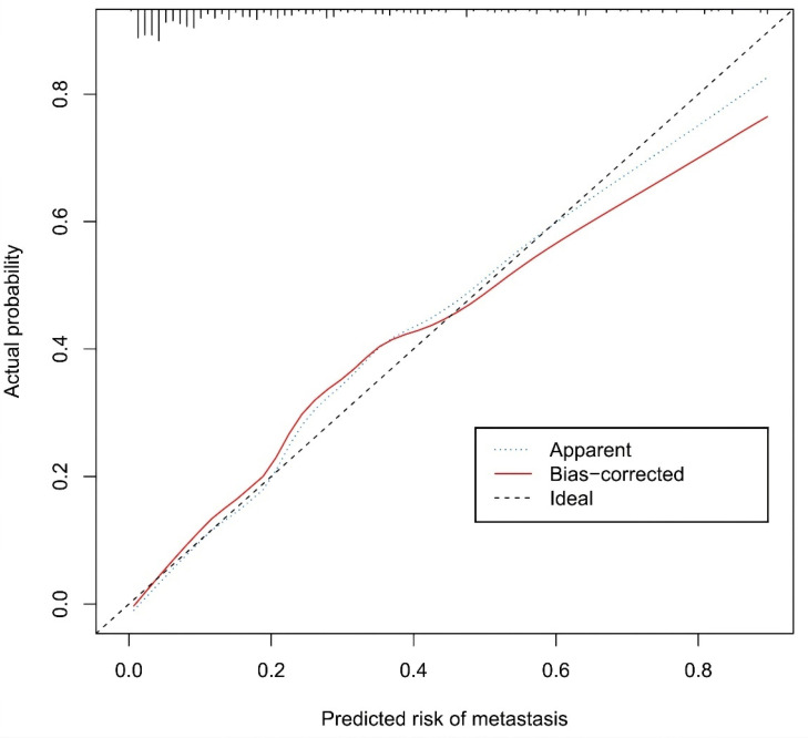
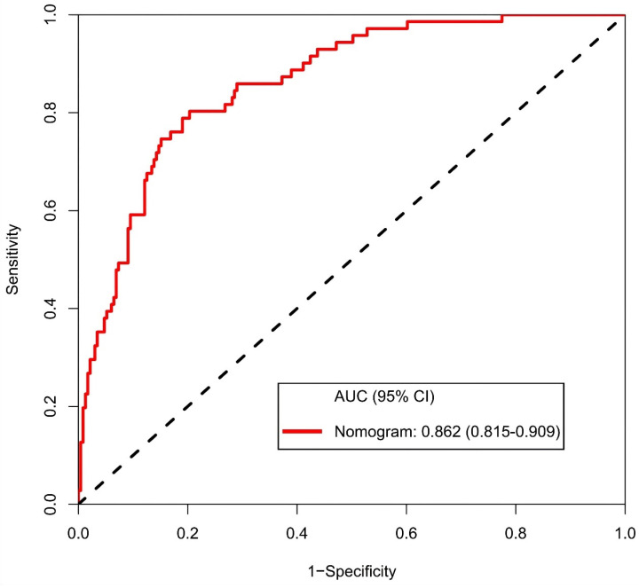
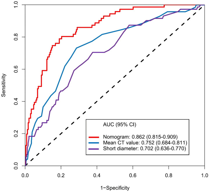
Preoperative assessment of axillary lymph node (ALN) status is essential for breast cancer treatment planning. This study prospectively analyzed risk factors for ALN metastasis by comparing high-resolution computed tomography (HRCT) imaging with pathology and developed a nomogram to aid in diagnosis. From April 2023 to May 2024, breast cancer patients confirmed by pathology participated in the study. All had chest HRCT before surgery, and ALN samples were anatomically matched to HRCT imaging and pathology. The least absolute shrinkage and selection operator (LASSO) regression helped refine metastasis features, and a nomogram was constructed using the final selected features determined by multivariate logistic regression. The nomogram's performance was evaluated with concordance index (C-index), calibration plot, and decision curve analysis, with internal validation through bootstrapping. A total of 302 ALN from 98 patients were included in this study. The predictors included in the nomogram encompassed the mean CT value, short diameter, border, and shape of ALN, as well as the Ki-67 status and histological grade of the primary tumor. The model exhibited satisfactory discrimination, with a C-index of 0.869 (95% CI: 0.826-0.912) and an AUC of 0.862 (95% CI, 0.815-0.909). The calibration curve demonstrated a high degree of concordance between the predicted and actual probabilities. The decision curve analysis demonstrated that the nomogram was clinically useful when the threshold for intervention was set at the metastasis possibility range of 1% to 86%. The nomogram combined with preoperative pathology and HRCT imaging have the potential to improve the evaluation of ALN status.

摘要

术前评估腋窝淋巴结(ALN)状态对于乳腺癌治疗方案的制定至关重要。本研究通过将高分辨率计算机断层扫描(HRCT)成像与病理学结果进行比较,前瞻性分析了ALN转移的危险因素,并开发了一种列线图辅助诊断。2023年4月至2024年5月,经病理确诊的乳腺癌患者参与了本研究。所有患者术前均进行了胸部HRCT检查,并且将ALN样本在解剖学上与HRCT成像及病理学结果进行匹配。最小绝对收缩和选择算子(LASSO)回归有助于优化转移特征,并使用多变量逻辑回归确定的最终选定特征构建列线图。使用一致性指数(C指数)、校准曲线和决策曲线分析评估列线图的性能,并通过自举法进行内部验证。本研究共纳入了98例患者的302个ALN。列线图中纳入的预测因素包括ALN的平均CT值、短径、边界和形态,以及原发肿瘤的Ki-67状态和组织学分级。该模型表现出令人满意的区分度,C指数为0.869(95%CI:0.826 - 0.912),曲线下面积(AUC)为0.862(95%CI,0.815 - 0.909)。校准曲线显示预测概率与实际概率之间具有高度一致性。决策曲线分析表明,当干预阈值设定在转移可能性范围为1%至86%时,列线图具有临床实用性。结合术前病理学和HRCT成像的列线图有潜力改善对ALN状态的评估。

https://cdn.ncbi.nlm.nih.gov/pmc/blobs/fe89/11493017/271eb88d1148/jcav15p6122g011.jpg
https://cdn.ncbi.nlm.nih.gov/pmc/blobs/fe89/11493017/34c5a052f90e/jcav15p6122g001.jpg
https://cdn.ncbi.nlm.nih.gov/pmc/blobs/fe89/11493017/b2c06d55f737/jcav15p6122g002.jpg
https://cdn.ncbi.nlm.nih.gov/pmc/blobs/fe89/11493017/6eccfefeb989/jcav15p6122g003.jpg
https://cdn.ncbi.nlm.nih.gov/pmc/blobs/fe89/11493017/7aec5d054e1d/jcav15p6122g004.jpg
https://cdn.ncbi.nlm.nih.gov/pmc/blobs/fe89/11493017/7bf94dd3cb8c/jcav15p6122g005.jpg
https://cdn.ncbi.nlm.nih.gov/pmc/blobs/fe89/11493017/0c0abdec09e8/jcav15p6122g006.jpg
https://cdn.ncbi.nlm.nih.gov/pmc/blobs/fe89/11493017/8cb14e32e49f/jcav15p6122g007.jpg
https://cdn.ncbi.nlm.nih.gov/pmc/blobs/fe89/11493017/ebe27cfb1e82/jcav15p6122g008.jpg
https://cdn.ncbi.nlm.nih.gov/pmc/blobs/fe89/11493017/3ab033b1394e/jcav15p6122g009.jpg
https://cdn.ncbi.nlm.nih.gov/pmc/blobs/fe89/11493017/5d2909e5a501/jcav15p6122g010.jpg
https://cdn.ncbi.nlm.nih.gov/pmc/blobs/fe89/11493017/271eb88d1148/jcav15p6122g011.jpg

相似文献

1
Development and Validation of a Nomogram for Axillary Lymph Node Metastasis Risk in Breast Cancer.乳腺癌腋窝淋巴结转移风险列线图的构建与验证
J Cancer. 2024 Oct 7;15(18):6122-6134. doi: 10.7150/jca.100651. eCollection 2024.
2
Development and Validation of a Nomogram for Predicting Axillary Lymph Node Metastasis in Breast Cancer.列线图预测乳腺癌腋窝淋巴结转移的建立与验证。
Clin Breast Cancer. 2023 Jul;23(5):538-545. doi: 10.1016/j.clbc.2023.04.002. Epub 2023 Apr 12.
3
Mammography-based radiomics nomogram: a potential biomarker to predict axillary lymph node metastasis in breast cancer.基于乳腺 X 线摄影的放射组学列线图:一种预测乳腺癌腋窝淋巴结转移的潜在生物标志物。
Br J Radiol. 2020 Jul;93(1111):20191019. doi: 10.1259/bjr.20191019. Epub 2020 May 27.
4
Dynamic contrast-enhanced MRI radiomics nomogram for predicting axillary lymph node metastasis in breast cancer.动态对比增强 MRI 放射组学列线图预测乳腺癌腋窝淋巴结转移。
Cancer Imaging. 2022 Apr 4;22(1):17. doi: 10.1186/s40644-022-00450-w.
5
Predicting the risk of axillary lymph node metastasis in early breast cancer patients based on ultrasonographic-clinicopathologic features and the use of nomograms: a prospective single-center observational study.基于超声临床病理特征和列线图模型预测早期乳腺癌患者腋窝淋巴结转移风险:一项前瞻性单中心观察性研究。
Eur Radiol. 2022 Dec;32(12):8200-8212. doi: 10.1007/s00330-022-08855-8. Epub 2022 Sep 28.
6
Nomogram for predicting axillary lymph node status after neoadjuvant chemotherapy in breast cancer.预测乳腺癌新辅助化疗后腋窝淋巴结状态的列线图
Transl Cancer Res. 2020 Nov;9(11):7054-7064. doi: 10.21037/tcr-20-2377.
7
Radiomics nomogram for predicting axillary lymph node metastasis in breast cancer based on DCE-MRI: A multicenter study.基于动态对比增强磁共振成像预测乳腺癌腋窝淋巴结转移的影像组学列线图:一项多中心研究
J Xray Sci Technol. 2023;31(2):247-263. doi: 10.3233/XST-221336.
8
Radiomics Nomogram of DCE-MRI for the Prediction of Axillary Lymph Node Metastasis in Breast Cancer.用于预测乳腺癌腋窝淋巴结转移的DCE-MRI影像组学列线图
Front Oncol. 2020 Oct 27;10:541849. doi: 10.3389/fonc.2020.541849. eCollection 2020.
9
Automated Breast Ultrasound (ABUS)-based radiomics nomogram: an individualized tool for predicting axillary lymph node tumor burden in patients with early breast cancer.基于自动乳腺超声(ABUS)的放射组学列线图:一种用于预测早期乳腺癌患者腋窝淋巴结肿瘤负担的个体化工具。
BMC Cancer. 2023 Apr 13;23(1):340. doi: 10.1186/s12885-023-10743-3.
10
Non-invasive prediction model of axillary lymph node status in patients with early-stage breast cancer: a feasibility study based on dynamic contrast-enhanced-MRI radiomics.基于动态对比增强 MRI 放射组学的早期乳腺癌患者腋窝淋巴结状态的无创预测模型:一项可行性研究。
Br J Radiol. 2024 Feb 2;97(1154):439-450. doi: 10.1093/bjr/tqad034.

引用本文的文献

1
Engineered upconversion nanoparticles for breast cancer theranostics.用于乳腺癌诊疗的工程化上转换纳米粒子
Theranostics. 2025 Jul 25;15(16):8259-8319. doi: 10.7150/thno.116153. eCollection 2025.
2
Development of a preoperative nomogram to identify low-risk early-stage breast cancer patients eligible for SLNB omission.开发一种术前列线图以识别适合省略前哨淋巴结活检的低风险早期乳腺癌患者。
World J Surg Oncol. 2025 Jul 7;23(1):268. doi: 10.1186/s12957-025-03921-z.

本文引用的文献

1
Can Hounsfield units on chest CT characterize breast nodules as cystic or solid?胸部CT上的亨氏单位能否将乳腺结节区分为囊性或实性?
Transl Breast Cancer Res. 2023 Nov 27;5:6. doi: 10.21037/tbcr-23-34. eCollection 2024.
2
Global cancer statistics 2022: GLOBOCAN estimates of incidence and mortality worldwide for 36 cancers in 185 countries.2022 年全球癌症统计数据:全球 185 个国家和地区 36 种癌症的发病率和死亡率全球估计数。
CA Cancer J Clin. 2024 May-Jun;74(3):229-263. doi: 10.3322/caac.21834. Epub 2024 Apr 4.
3
[Application value of CT examination of lymph node short diameter in evaluating cardia-left gastric lymph node metastasis in thoracic esophageal squamous cell carcinoma].
[CT检查淋巴结短径在评估胸段食管鳞状细胞癌贲门-胃左淋巴结转移中的应用价值]
Zhonghua Zhong Liu Za Zhi. 2023 Nov 23;45(11):962-966. doi: 10.3760/cma.j.cn112152-20220313-00173.
4
The prognostic value of the histological shape of tumor negative sentinel nodes in breast cancer.肿瘤阴性前哨淋巴结组织学形态的预后价值在乳腺癌中的体现。
Front Immunol. 2023 Oct 30;14:1258641. doi: 10.3389/fimmu.2023.1258641. eCollection 2023.
5
Construction of a comprehensive predictive model for axillary lymph node metastasis in breast cancer: a retrospective study.构建乳腺癌腋窝淋巴结转移的综合预测模型:一项回顾性研究。
BMC Cancer. 2023 Oct 24;23(1):1028. doi: 10.1186/s12885-023-11498-7.
6
Lymph node metastases in breast cancer: Mechanisms and molecular imaging.乳腺癌淋巴结转移:机制与分子影像学。
Clin Imaging. 2023 Nov;103:109985. doi: 10.1016/j.clinimag.2023.109985. Epub 2023 Sep 21.
7
Dynamic CD8 T cell responses to cancer immunotherapy in human regional lymph nodes are disrupted in metastatic lymph nodes.在转移性淋巴结中,癌症免疫治疗在人体区域淋巴结中诱导的动态 CD8 T 细胞反应被打乱。
Cell. 2023 Mar 16;186(6):1127-1143.e18. doi: 10.1016/j.cell.2023.02.021.
8
Diagnostic Usefulness of Diffusion-Weighted MRI for Axillary Lymph Node Evaluation in Patients with Breast Cancer.扩散加权磁共振成像在乳腺癌患者腋窝淋巴结评估中的诊断价值
Diagnostics (Basel). 2023 Jan 31;13(3):513. doi: 10.3390/diagnostics13030513.
9
Assessing Whether Morphological Changes in Axillary Lymph Node Have Already Occurred Prior to Metastasis in Breast Cancer Patients by Ultrasound.超声评估乳腺癌患者腋窝淋巴结转移前的形态学变化。
Medicina (Kaunas). 2022 Nov 18;58(11):1674. doi: 10.3390/medicina58111674.
10
Chest CT for Breast Cancer Diagnosis.用于乳腺癌诊断的胸部CT
Life (Basel). 2022 Oct 26;12(11):1699. doi: 10.3390/life12111699.