• 文献检索
  • 文档翻译
  • 深度研究
  • 学术资讯
  • Suppr Zotero 插件Zotero 插件
  • 邀请有礼
  • 套餐&价格
  • 历史记录
应用&插件
Suppr Zotero 插件Zotero 插件浏览器插件Mac 客户端Windows 客户端微信小程序
定价
高级版会员购买积分包购买API积分包
服务
文献检索文档翻译深度研究API 文档MCP 服务
关于我们
关于 Suppr公司介绍联系我们用户协议隐私条款
关注我们

Suppr 超能文献

核心技术专利:CN118964589B侵权必究
粤ICP备2023148730 号-1Suppr @ 2026

文献检索

告别复杂PubMed语法,用中文像聊天一样搜索,搜遍4000万医学文献。AI智能推荐,让科研检索更轻松。

立即免费搜索

文件翻译

保留排版,准确专业,支持PDF/Word/PPT等文件格式,支持 12+语言互译。

免费翻译文档

深度研究

AI帮你快速写综述,25分钟生成高质量综述,智能提取关键信息,辅助科研写作。

立即免费体验

基于乳腺 X 线摄影的放射组学列线图:一种预测乳腺癌腋窝淋巴结转移的潜在生物标志物。

Mammography-based radiomics nomogram: a potential biomarker to predict axillary lymph node metastasis in breast cancer.

机构信息

Department of Radiology, Henan Provincial People's Hospital & Imaging Diagnosis of Neurological Diseases and Research Laboratory of Henan Province & People's Hospital of Zhengzhou University, Henan, China, 450003.

Department of Hematology, Henan Provincial People's Hospital & People's Hospital of Zhengzhou University, Henan, China, 450003.

出版信息

Br J Radiol. 2020 Jul;93(1111):20191019. doi: 10.1259/bjr.20191019. Epub 2020 May 27.

DOI:10.1259/bjr.20191019
PMID:32401540
原文链接:https://pmc.ncbi.nlm.nih.gov/articles/PMC7336077/
Abstract

OBJECTIVE

To establish a radiomics nomogram by integrating clinical risk factors and radiomics features extracted from digital mammography (MG) images for pre-operative prediction of axillary lymph node (ALN) metastasis in breast cancer.

METHODS

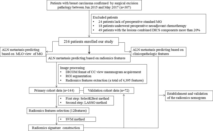
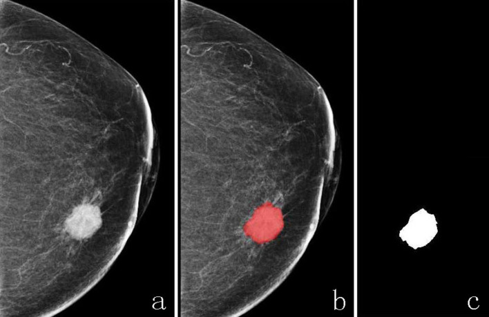
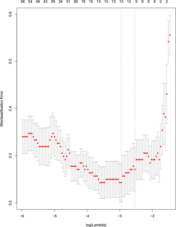
216 patients with breast cancer lesions confirmed by surgical excision pathology were divided into the primary cohort ( = 144) and validation cohort ( = 72). Radiomics features were extracted from craniocaudal (CC) view of mammograms, and radiomics features selection were performed using the methods of ANOVA F-value and least absolute shrinkage and selection operator; then a radiomics signature was constructed with the method of support vector machine. Multivariate logistic regression analysis was used to establish a radiomics nomogram based on the combination of radiomics signature and clinical factors. The C-index and calibration curves were derived based on the regression analysis both in the primary and validation cohorts.

RESULTS

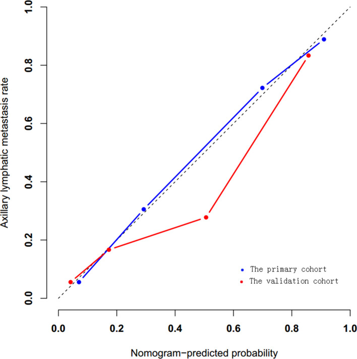
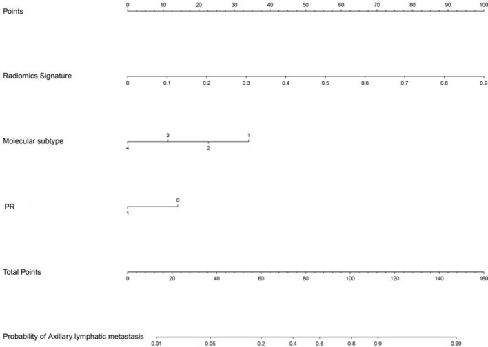
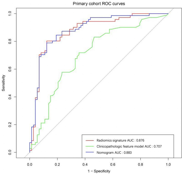
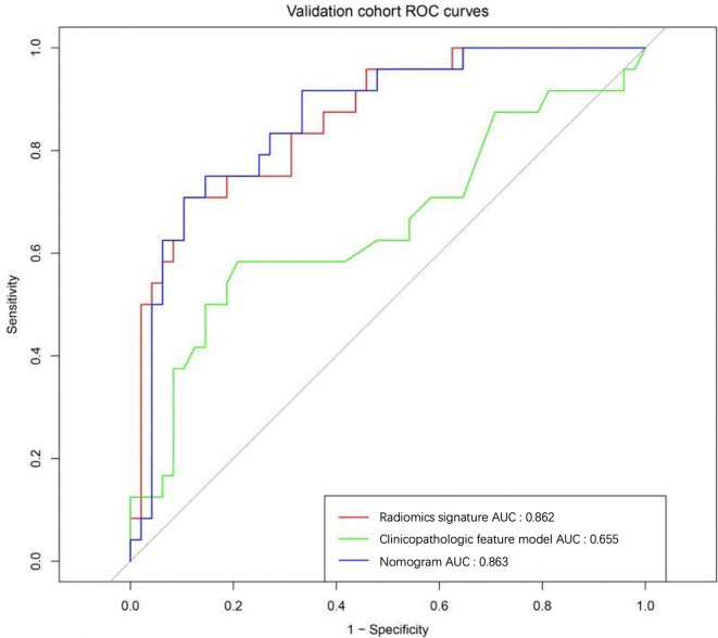
95 of 216 patients were confirmed with ALN metastasis by pathology, and 52 cases were diagnosed as ALN metastasis based on MG-reported criteria. The sensitivity, specificity, accuracy and AUC (area under the receiver operating characteristic curve of MG-reported criteria were 42.7%, 90.8%, 24.1% and 0.666 (95% confidence interval: 0.591-0.741]. The radiomics nomogram, comprising progesterone receptor status, molecular subtype and radiomics signature, showed good calibration and better favorite performance for the metastatic ALN detection (AUC 0.883 and 0.863 in the primary and validation cohorts) than each independent clinical features (AUC 0.707 and 0.657 in the primary and validation cohorts) and radiomics signature (AUC 0.876 and 0.862 in the primary and validation cohorts).

CONCLUSION

The MG-based radiomics nomogram could be used as a non-invasive and reliable tool in predicting ALN metastasis and may facilitate to assist clinicians for pre-operative decision-making.

ADVANCES IN KNOWLEDGE

ALN status remains among the most important breast cancer prognostic factors and is essential for making treatment decisions. However, the value of detecting metastatic ALN by MG is very limited. The studies on pre-operative ALN metastasis prediction using the method of MG-based radiomics in breast cancer are very few. Therefore, we studied whether MG-based radiomics nomogram could be used as a predictive biomarker for the detection of metastatic ALN.

摘要

目的

通过整合临床风险因素和从数字乳腺 X 线摄影(MG)图像中提取的放射组学特征,建立术前预测乳腺癌腋窝淋巴结(ALN)转移的放射组学列线图。

方法

将 216 例经手术切除病理证实的乳腺癌病变患者分为主队列(n=144)和验证队列(n=72)。从乳腺头尾位(CC)视图的 MG 图像中提取放射组学特征,并采用方差分析 F 值和最小绝对值收缩和选择算子(least absolute shrinkage and selection operator)的方法进行放射组学特征选择;然后使用支持向量机(support vector machine)的方法构建放射组学特征模型。采用多变量逻辑回归分析,基于放射组学特征和临床因素建立放射组学列线图。基于回归分析,在主队列和验证队列中分别获得 C 指数和校准曲线。

结果

216 例患者中,95 例经病理证实存在 ALN 转移,52 例经 MG 报告标准诊断为 ALN 转移。MG 报告标准的敏感性、特异性、准确性和 AUC(接收器工作特征曲线下面积)分别为 42.7%、90.8%、24.1%和 0.666(95%置信区间:0.591-0.741]。包括孕激素受体状态、分子亚型和放射组学特征的放射组学列线图,对转移性 ALN 检测具有良好的校准和更好的优势表现(主队列和验证队列的 AUC 分别为 0.883 和 0.863),优于每个独立的临床特征(主队列和验证队列的 AUC 分别为 0.707 和 0.657)和放射组学特征(主队列和验证队列的 AUC 分别为 0.876 和 0.862)。

结论

基于 MG 的放射组学列线图可作为预测 ALN 转移的一种非侵入性、可靠的工具,并有助于辅助临床医生进行术前决策。

知识进展

ALN 状态仍然是乳腺癌最重要的预后因素之一,对于治疗决策至关重要。然而,MG 检测转移性 ALN 的价值非常有限。在乳腺癌中,使用 MG 基于放射组学的方法进行术前 ALN 转移预测的研究非常少。因此,我们研究了 MG 基于放射组学的列线图是否可作为检测转移性 ALN 的预测生物标志物。

https://cdn.ncbi.nlm.nih.gov/pmc/blobs/bc6e/7336077/5bd7c4a47403/bjr.20191019.g008.jpg
https://cdn.ncbi.nlm.nih.gov/pmc/blobs/bc6e/7336077/b2d0638e78d1/bjr.20191019.g001.jpg
https://cdn.ncbi.nlm.nih.gov/pmc/blobs/bc6e/7336077/b809dd0b9713/bjr.20191019.g002.jpg
https://cdn.ncbi.nlm.nih.gov/pmc/blobs/bc6e/7336077/a2692aaa339c/bjr.20191019.g003.jpg
https://cdn.ncbi.nlm.nih.gov/pmc/blobs/bc6e/7336077/df36ddc84aa0/bjr.20191019.g004.jpg
https://cdn.ncbi.nlm.nih.gov/pmc/blobs/bc6e/7336077/5d98019e6439/bjr.20191019.g005.jpg
https://cdn.ncbi.nlm.nih.gov/pmc/blobs/bc6e/7336077/4bd165f2fd21/bjr.20191019.g006.jpg
https://cdn.ncbi.nlm.nih.gov/pmc/blobs/bc6e/7336077/f23ed3e8654f/bjr.20191019.g007.jpg
https://cdn.ncbi.nlm.nih.gov/pmc/blobs/bc6e/7336077/5bd7c4a47403/bjr.20191019.g008.jpg

相似文献

1
Mammography-based radiomics nomogram: a potential biomarker to predict axillary lymph node metastasis in breast cancer.基于乳腺 X 线摄影的放射组学列线图:一种预测乳腺癌腋窝淋巴结转移的潜在生物标志物。
Br J Radiol. 2020 Jul;93(1111):20191019. doi: 10.1259/bjr.20191019. Epub 2020 May 27.
2
Ultrasound-based radiomics nomogram: A potential biomarker to predict axillary lymph node metastasis in early-stage invasive breast cancer.基于超声的放射组学列线图:预测早期浸润性乳腺癌腋窝淋巴结转移的潜在生物标志物。
Eur J Radiol. 2019 Oct;119:108658. doi: 10.1016/j.ejrad.2019.108658. Epub 2019 Sep 7.
3
Dynamic contrast-enhanced MRI radiomics nomogram for predicting axillary lymph node metastasis in breast cancer.动态对比增强 MRI 放射组学列线图预测乳腺癌腋窝淋巴结转移。
Cancer Imaging. 2022 Apr 4;22(1):17. doi: 10.1186/s40644-022-00450-w.
4
A two-center study of a combined nomogram based on mammography and MRI to predict ALN metastasis in breast cancer.基于乳腺 X 线摄影和 MRI 的联合列线图预测乳腺癌前哨淋巴结转移的两中心研究。
Magn Reson Imaging. 2024 Jul;110:128-137. doi: 10.1016/j.mri.2024.04.019. Epub 2024 Apr 15.
5
Radiomics nomogram of contrast-enhanced spectral mammography for prediction of axillary lymph node metastasis in breast cancer: a multicenter study.对比增强光谱乳腺摄影放射组学列线图预测乳腺癌腋窝淋巴结转移:一项多中心研究。
Eur Radiol. 2020 Dec;30(12):6732-6739. doi: 10.1007/s00330-020-07016-z. Epub 2020 Jun 30.
6
Could Ultrasound-Based Radiomics Noninvasively Predict Axillary Lymph Node Metastasis in Breast Cancer?基于超声的影像组学能否无创预测乳腺癌腋窝淋巴结转移?
J Ultrasound Med. 2020 Oct;39(10):1897-1905. doi: 10.1002/jum.15294. Epub 2020 Apr 24.
7
Magnetic resonance imaging radiomics predicts preoperative axillary lymph node metastasis to support surgical decisions and is associated with tumor microenvironment in invasive breast cancer: A machine learning, multicenter study.磁共振成像放射组学预测术前腋窝淋巴结转移以支持手术决策,并与浸润性乳腺癌的肿瘤微环境相关:一项机器学习、多中心研究。
EBioMedicine. 2021 Jul;69:103460. doi: 10.1016/j.ebiom.2021.103460. Epub 2021 Jul 4.
8
Preoperative Prediction of Axillary Lymph Node Metastasis in Breast Cancer Using Mammography-Based Radiomics Method.基于乳腺 X 线摄影的放射组学方法预测乳腺癌腋窝淋巴结转移
Sci Rep. 2019 Mar 14;9(1):4429. doi: 10.1038/s41598-019-40831-z.
9
Radiomic Nomogram for Predicting Axillary Lymph Node Metastasis in Patients with Breast Cancer.基于影像组学的 nomogram 模型预测乳腺癌患者腋窝淋巴结转移
Acad Radiol. 2024 Mar;31(3):788-799. doi: 10.1016/j.acra.2023.10.026. Epub 2023 Nov 4.
10
Non-invasive prediction model of axillary lymph node status in patients with early-stage breast cancer: a feasibility study based on dynamic contrast-enhanced-MRI radiomics.基于动态对比增强 MRI 放射组学的早期乳腺癌患者腋窝淋巴结状态的无创预测模型:一项可行性研究。
Br J Radiol. 2024 Feb 2;97(1154):439-450. doi: 10.1093/bjr/tqad034.

引用本文的文献

1
Deep learning on routine full-breast mammograms enhances lymph node metastasis prediction in early breast cancer.基于常规全乳钼靶X线摄影的深度学习可提高早期乳腺癌淋巴结转移的预测能力。
NPJ Digit Med. 2025 Jul 10;8(1):425. doi: 10.1038/s41746-025-01831-8.
2
Clinical Significance of the Serum 3'tRF-AlaAGC, Neutrophil to High-Density Lipoprotein Ratio, and Lymphocyte-to-Monocyte Ratio in Breast Cancer with Lymph Node Metastasis.血清3'tRF-AlaAGC、中性粒细胞与高密度脂蛋白比值及淋巴细胞与单核细胞比值在伴淋巴结转移乳腺癌中的临床意义
J Inflamm Res. 2025 May 7;18:5979-5991. doi: 10.2147/JIR.S518232. eCollection 2025.
3
Enhancing Specificity in Predicting Axillary Lymph Node Metastasis in Breast Cancer through an Interpretable Machine Learning Model with CEM and Ultrasound Integration.

本文引用的文献

1
Multiparametric MRI-Based Radiomics Nomogram for Predicting Lymph Node Metastasis in Early-Stage Cervical Cancer.基于多参数磁共振成像的影像组学列线图预测早期宫颈癌淋巴结转移
J Magn Reson Imaging. 2020 Sep;52(3):885-896. doi: 10.1002/jmri.27101. Epub 2020 Feb 25.
2
Ultrasound-based radiomics nomogram: A potential biomarker to predict axillary lymph node metastasis in early-stage invasive breast cancer.基于超声的放射组学列线图:预测早期浸润性乳腺癌腋窝淋巴结转移的潜在生物标志物。
Eur J Radiol. 2019 Oct;119:108658. doi: 10.1016/j.ejrad.2019.108658. Epub 2019 Sep 7.
3
CT radiomics nomogram for the preoperative prediction of lymph node metastasis in gastric cancer.
通过具有CEM和超声整合的可解释机器学习模型提高乳腺癌腋窝淋巴结转移预测的特异性
Technol Cancer Res Treat. 2025 Jan-Dec;24:15330338251334735. doi: 10.1177/15330338251334735. Epub 2025 Apr 17.
4
Preoperative prediction of nodal status using clinical data and artificial intelligence derived mammogram features enabling abstention of sentinel lymph node biopsy in breast cancer.利用临床数据和人工智能提取的乳房X光检查特征进行术前淋巴结状态预测,可避免乳腺癌患者进行前哨淋巴结活检。
Front Oncol. 2024 Jul 10;14:1394448. doi: 10.3389/fonc.2024.1394448. eCollection 2024.
5
Assessment of the axilla in women with early-stage breast cancer undergoing primary surgery: a review.早期乳腺癌行原发性手术的女性腋窝评估:综述。
World J Surg Oncol. 2024 May 9;22(1):127. doi: 10.1186/s12957-024-03394-6.
6
Radiomics nomogram for predicting axillary lymph node metastasis-a potential method to address the limitation of axilla coverage in cone-beam breast CT: a bi-center retrospective study.基于放射组学的腋窝淋巴结转移预测列线图:一种解决锥形束乳腺 CT 腋窝覆盖不足的潜在方法:一项双中心回顾性研究。
Radiol Med. 2023 Dec;128(12):1472-1482. doi: 10.1007/s11547-023-01731-5. Epub 2023 Oct 19.
7
Radiomics in cone-beam breast CT for the prediction of axillary lymph node metastasis in breast cancer: a multi-center multi-device study.锥形束乳腺 CT 中的放射组学用于预测乳腺癌腋窝淋巴结转移:一项多中心多设备研究。
Eur Radiol. 2024 Apr;34(4):2576-2589. doi: 10.1007/s00330-023-10256-4. Epub 2023 Oct 2.
8
Characterization of Breast Tumors from MR Images Using Radiomics and Machine Learning Approaches.使用影像组学和机器学习方法对乳腺肿瘤进行特征分析。
J Pers Med. 2023 Jun 28;13(7):1062. doi: 10.3390/jpm13071062.
9
Hype or hope? A review of challenges in balancing tumor control and treatment toxicity in breast cancer from the perspective of the radiation oncologist.炒作还是希望?从放射肿瘤学家的角度看乳腺癌肿瘤控制与治疗毒性平衡的挑战。
Clin Transl Oncol. 2024 Mar;26(3):561-573. doi: 10.1007/s12094-023-03287-2. Epub 2023 Jul 28.
10
Use of machine learning with two-dimensional synthetic mammography for axillary lymph node metastasis prediction in breast cancer: a preliminary study.二维合成乳腺钼靶联合机器学习在乳腺癌腋窝淋巴结转移预测中的应用:一项初步研究。
Transl Cancer Res. 2023 May 31;12(5):1232-1240. doi: 10.21037/tcr-22-2668. Epub 2023 Apr 28.
CT 放射组学列线图预测胃癌淋巴结转移的术前评估。
Eur Radiol. 2020 Feb;30(2):976-986. doi: 10.1007/s00330-019-06398-z. Epub 2019 Aug 29.
4
Preoperative prediction of pelvic lymph nodes metastasis in early-stage cervical cancer using radiomics nomogram developed based on T2-weighted MRI and diffusion-weighted imaging.基于 T2 加权 MRI 和弥散加权成像的放射组学列线图术前预测早期宫颈癌盆腔淋巴结转移
Eur J Radiol. 2019 May;114:128-135. doi: 10.1016/j.ejrad.2019.01.003. Epub 2019 Mar 20.
5
Preoperative Prediction of Axillary Lymph Node Metastasis in Breast Cancer Using Mammography-Based Radiomics Method.基于乳腺 X 线摄影的放射组学方法预测乳腺癌腋窝淋巴结转移
Sci Rep. 2019 Mar 14;9(1):4429. doi: 10.1038/s41598-019-40831-z.
6
Differentiating axillary lymph node metastasis in invasive breast cancer patients: A comparison of radiomic signatures from multiparametric breast MR sequences.鉴别浸润性乳腺癌患者腋窝淋巴结转移:多参数乳腺磁共振序列的放射组学特征比较。
J Magn Reson Imaging. 2019 Oct;50(4):1125-1132. doi: 10.1002/jmri.26701. Epub 2019 Mar 7.
7
Preoperative Prediction of Axillary Lymph Node Metastasis in Breast Cancer using Radiomics Features of DCE-MRI.基于 DCE-MRI 影像组学特征预测乳腺癌腋窝淋巴结转移
Sci Rep. 2019 Feb 19;9(1):2240. doi: 10.1038/s41598-019-38502-0.
8
Radiomic nomogram for prediction of axillary lymph node metastasis in breast cancer.基于影像组学的乳腺癌腋窝淋巴结转移预测列线图
Eur Radiol. 2019 Jul;29(7):3820-3829. doi: 10.1007/s00330-018-5981-2. Epub 2019 Jan 30.
9
Organotropism: new insights into molecular mechanisms of breast cancer metastasis.器官嗜性:乳腺癌转移分子机制的新见解
NPJ Precis Oncol. 2018 Feb 16;2(1):4. doi: 10.1038/s41698-018-0047-0. eCollection 2018.
10
A new approach to predict lymph node metastasis in solid lung adenocarcinoma: a radiomics nomogram.预测实性肺腺癌淋巴结转移的新方法:一种影像组学列线图
J Thorac Dis. 2018 Apr;10(Suppl 7):S807-S819. doi: 10.21037/jtd.2018.03.126.