Zhang Peisen, Li Yao, Li Xiaoqi, Wang Yudong, Lin Hua, Zhang Ni, Li Wenyue, Jing Lihong, Jiao Mingxia, Luo Xiliang, Hou Yi
Key Laboratory of Optic-Electric Sensing and Analytical Chemistry for Life Science, MOE, College of Chemistry and Molecular Engineering, Qingdao University of Science and Technology, Qingdao, 266042, China.
College of Life Science and Technology, Beijing University of Chemical Technology, Beijing, 100029, China.
J Nanobiotechnology. 2024 Dec 18;22(1):757. doi: 10.1186/s12951-024-03042-x.
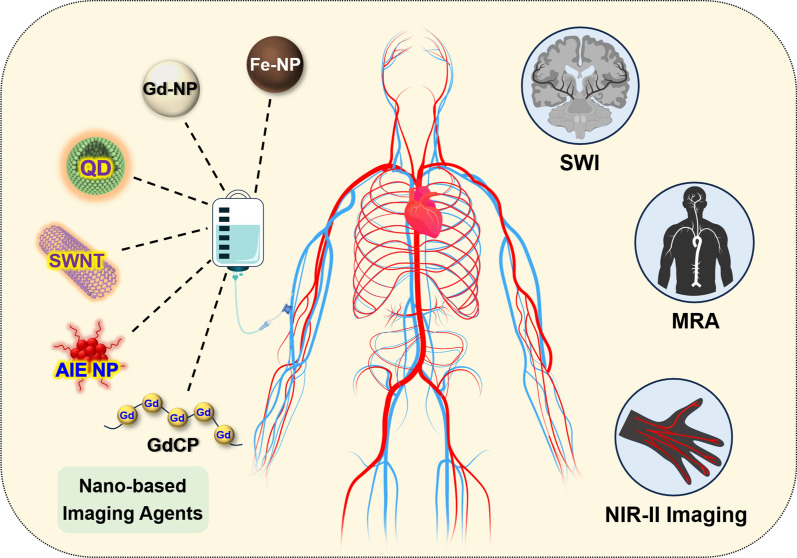
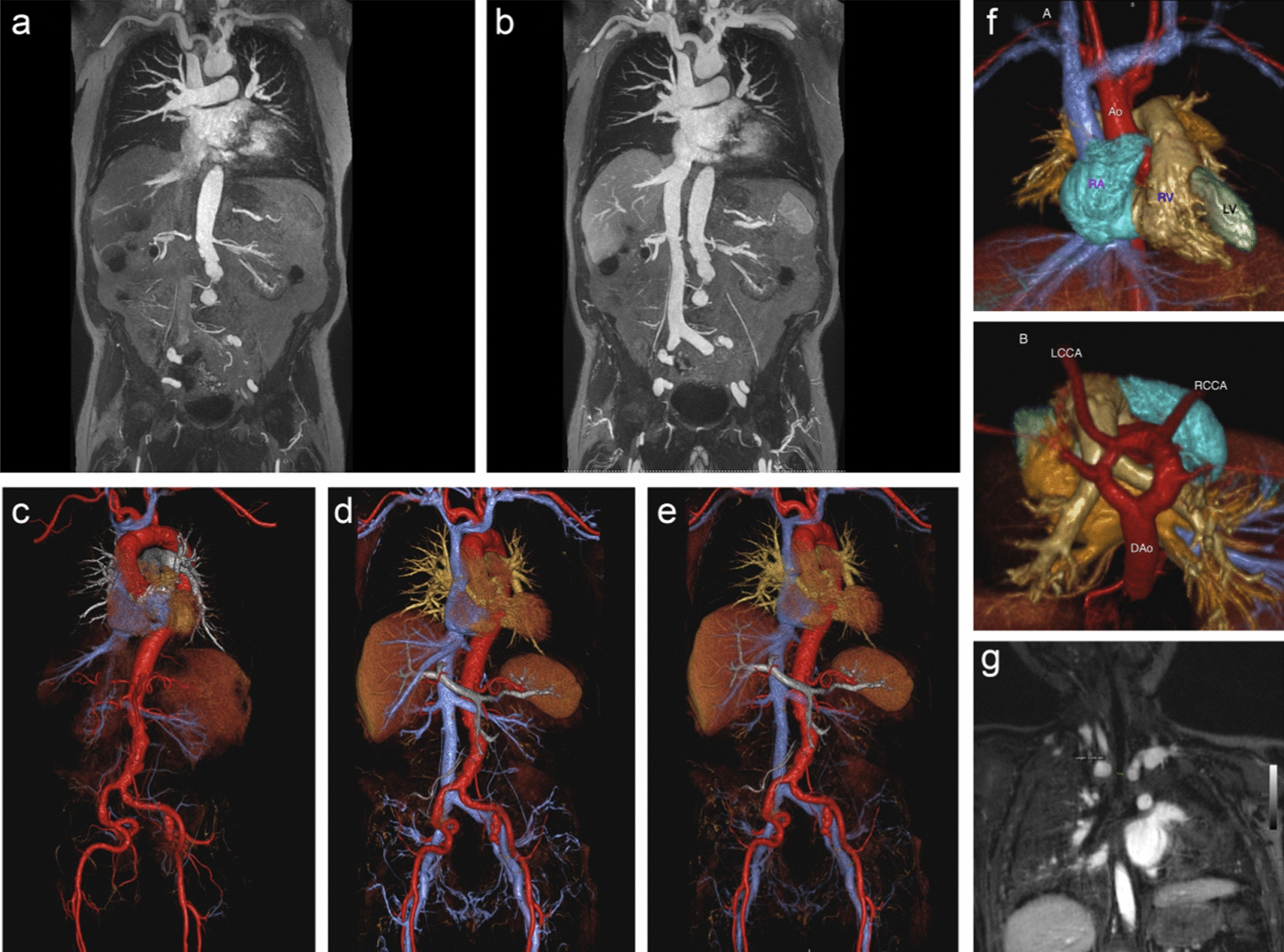
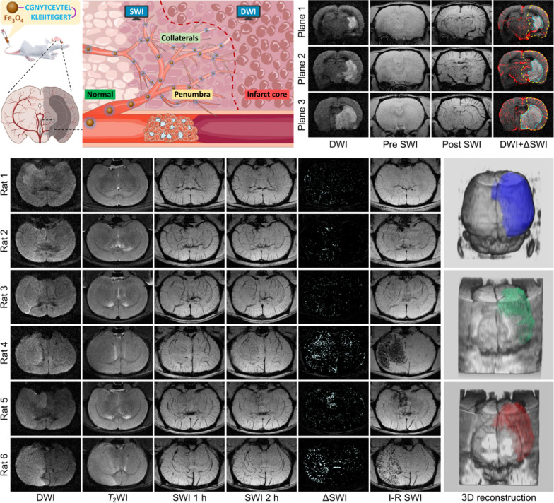
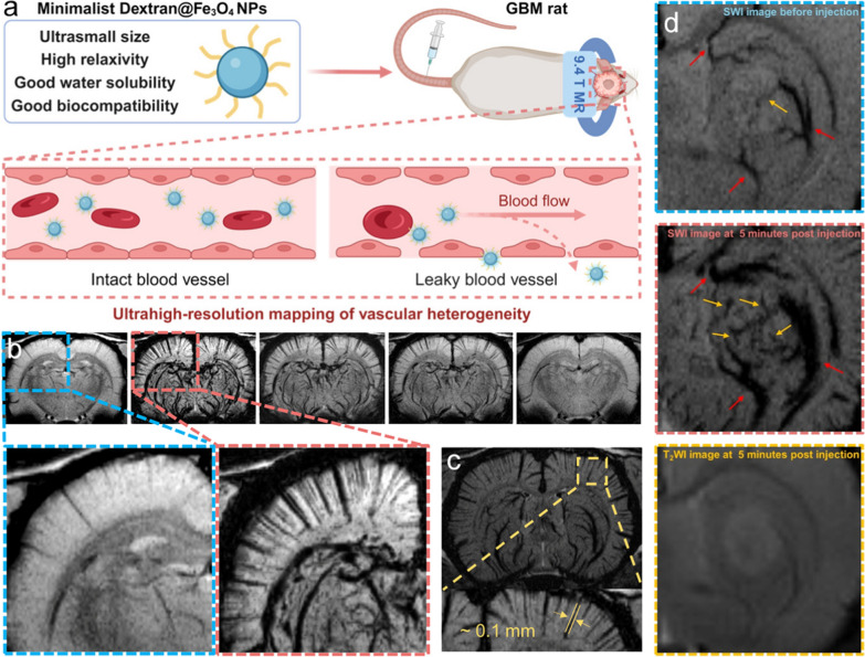
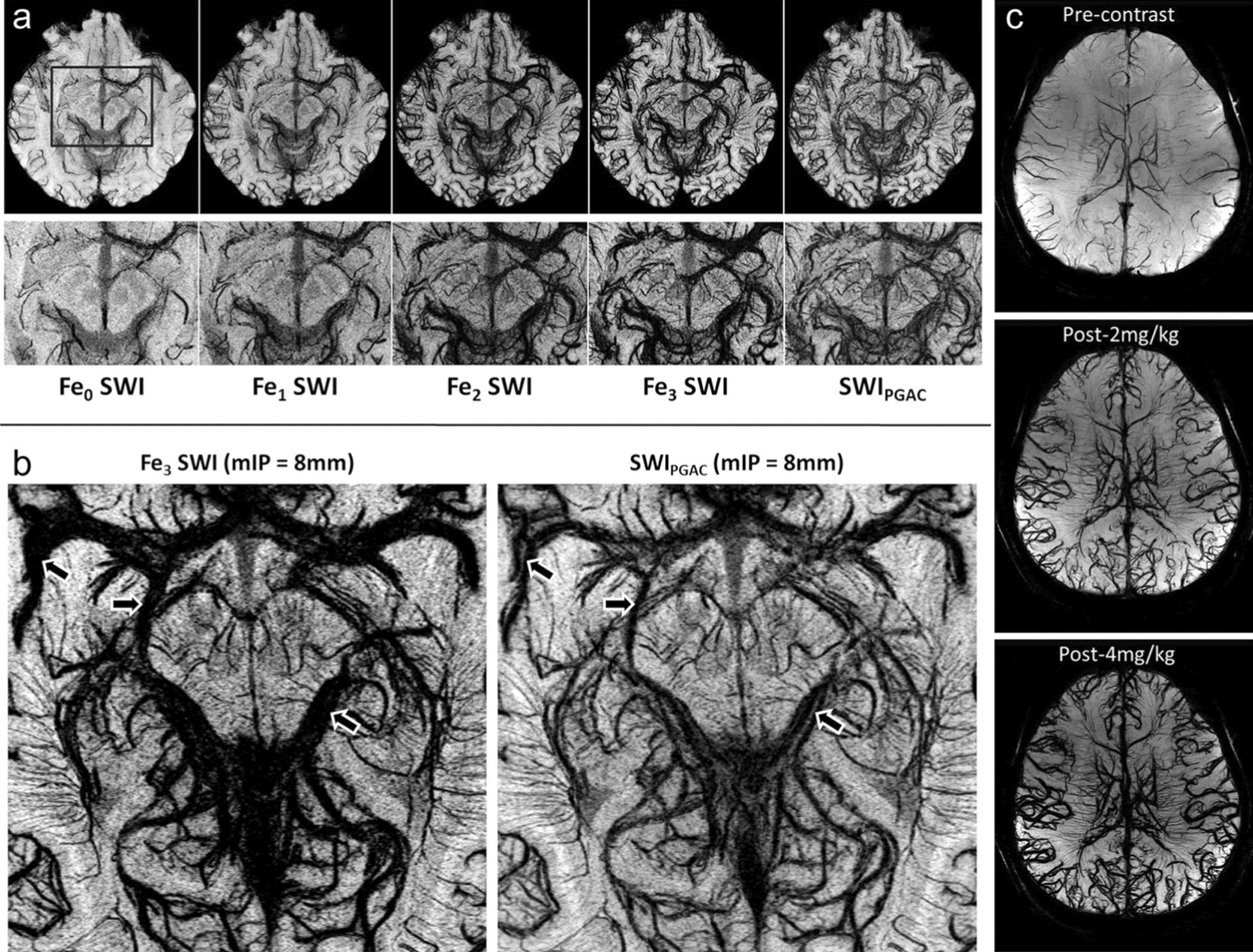
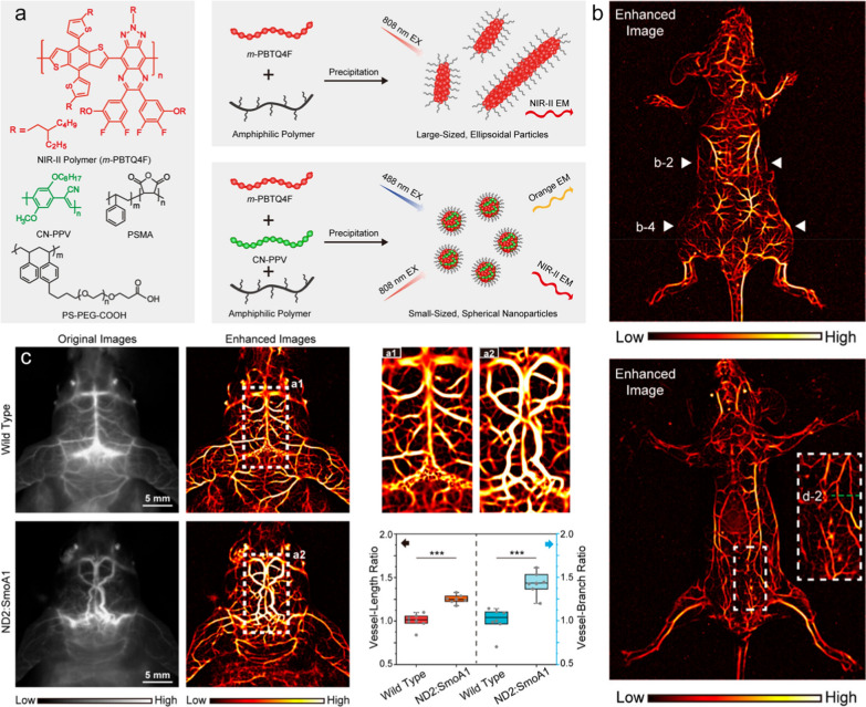
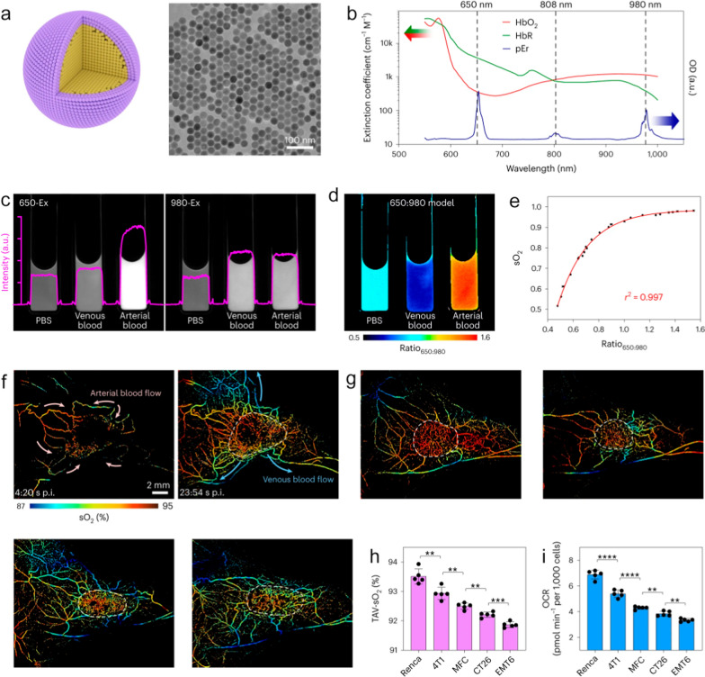
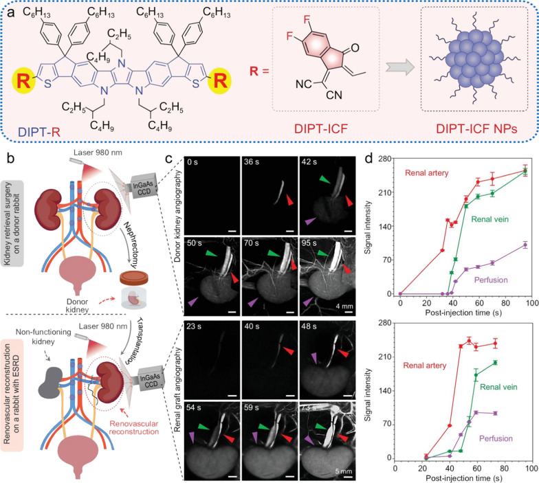
Vascular dysfunction, characterized by changes in anatomy, hemodynamics, and molecular expressions of vasculatures, is closely linked to the onset and development of diseases, emphasizing the importance of its detection. In clinical practice, medical imaging has been utilized as a significant tool in the assessment of vascular dysfunction, however, traditional imaging techniques still lack sufficient resolution for visualizing the complex microvascular systems. Over the past decade, with the rapid advancement of nanotechnology and the emergence of corresponding detection facilities, engineered nanomaterials offer new alternatives to traditional contrast agents. Compared with conventional small molecule counterparts, nanomaterials possess numerous advantages for vascular imaging, holding the potential to significantly advance related technologies. In this review, the latest developments in nanotechnology-assisted vascular imaging research across different imaging modalities, including contrast-enhanced magnetic resonance (MR) angiography, susceptibility-weighted imaging (SWI), and fluorescence imaging in the second near-infrared window (NIR-II) are summarized. Additionally, the advancements of preclinical and clinical studies related to these nanotechnology-enhanced vascular imaging approaches are outlined, with subsequent discussion on the current challenges and future prospects in both basic research and clinical translation.
血管功能障碍以血管的解剖结构、血流动力学和分子表达变化为特征,与疾病的发生和发展密切相关,这凸显了检测血管功能障碍的重要性。在临床实践中,医学成像已被用作评估血管功能障碍的重要工具,然而,传统成像技术在可视化复杂微血管系统方面仍缺乏足够的分辨率。在过去十年中,随着纳米技术的迅速发展和相应检测设备的出现,工程纳米材料为传统造影剂提供了新的替代品。与传统小分子造影剂相比,纳米材料在血管成像方面具有众多优势,有望显著推动相关技术发展。在本综述中,总结了纳米技术辅助血管成像研究在不同成像模式下的最新进展,包括对比增强磁共振(MR)血管造影、磁敏感加权成像(SWI)和第二近红外窗口(NIR-II)荧光成像。此外,还概述了与这些纳米技术增强血管成像方法相关的临床前和临床研究进展,随后讨论了基础研究和临床转化中当前面临的挑战和未来前景。