• 文献检索
  • 文档翻译
  • 深度研究
  • 学术资讯
  • Suppr Zotero 插件Zotero 插件
  • 邀请有礼
  • 套餐&价格
  • 历史记录
应用&插件
Suppr Zotero 插件Zotero 插件浏览器插件Mac 客户端Windows 客户端微信小程序
定价
高级版会员购买积分包购买API积分包
服务
文献检索文档翻译深度研究API 文档MCP 服务
关于我们
关于 Suppr公司介绍联系我们用户协议隐私条款
关注我们

Suppr 超能文献

核心技术专利:CN118964589B侵权必究
粤ICP备2023148730 号-1Suppr @ 2026

文献检索

告别复杂PubMed语法,用中文像聊天一样搜索,搜遍4000万医学文献。AI智能推荐,让科研检索更轻松。

立即免费搜索

文件翻译

保留排版,准确专业,支持PDF/Word/PPT等文件格式,支持 12+语言互译。

免费翻译文档

深度研究

AI帮你快速写综述,25分钟生成高质量综述,智能提取关键信息,辅助科研写作。

立即免费体验

实时三维经胸超声心动图节段容积分析:一种评估缺血性心脏病患者左心室壁节段运动的定量且客观的工具。

Real-time three-dimensional transthoracic echocardiographic segmental volume analysis: a quantitative and objective tool for assessing regional left ventricle wall motion in patients with ischemic heart disease.

作者信息

Kwak Jin-Hwan, Choi Kang-Un, Park Jong-Il, Nam Jong-Ho, Lee Chan-Hee, Kim Ung, Park Jong-Seon, Son Jang-Won

机构信息

Division of Cardiology, Department of Internal Medicine, Yeungnam University Medical Center, Daegu, Republic of Korea.

出版信息

J Cardiovasc Imaging. 2024 Dec 23;32(1):40. doi: 10.1186/s44348-024-00040-3.

DOI:10.1186/s44348-024-00040-3
PMID:39716242
原文链接:https://pmc.ncbi.nlm.nih.gov/articles/PMC11665206/
Abstract

BACKGROUND

Evaluation of regional left ventricle function using two-dimensional echocardiography (2DE) in patients with ischemic heart disease has limitations due to its low objectivity and qualitative nature. In addition, 2DE is limited because multiple acoustic windows are used to obtain the image, whereas three-dimensional echocardiography (3DE) uses a single window. This study aims to demonstrate the clinical utility of 3DE segmental volume analysis for evaluating regional wall motion abnormality (RWMA).

METHODS

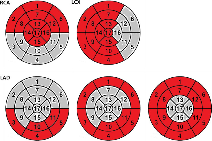
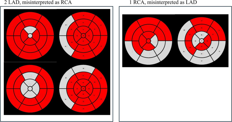
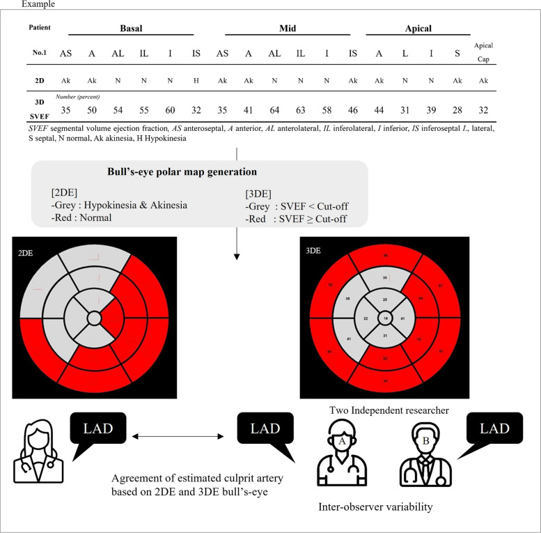
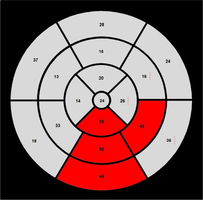
This retrospective study included 33 patients with ischemic heart disease and single-vessel territory RWMA confirmed on coronary angiography. RWMA was visually assessed using 2DE, generating 17-segment bull's-eye polar maps, and 3DE. In the 3DE study, two independent observers analyzed segmental volumes and segmental volume ejection fractions (SVEFs) using QLAB 3D quantification software. The optimal SVEF cutoff value differentiating normal from abnormal was determined using receiver operating curve analysis. The accuracy of 3DE in predicting culprit coronary arteries was compared with that of 2DE using Cohen κ coefficients, which also were used for interobserver and intraobserver variability assessments.

RESULTS

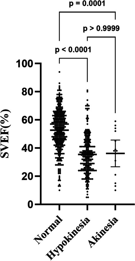
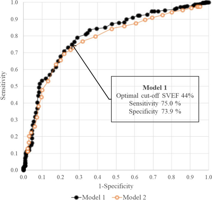
Mean 3DE SVEFs were significantly lower in segments showing RWMA on 2DE. The optimal SVEF cutoff value was 44%, with sensitivity of 75.0% and specificity of 73.9% (area under the curve, 0.801; 95% CI, 0.763-0.838; P < 0.001). The reliability of 3DE-derived bull's-eye predictions of culprit coronary arteries was 81.8% (κ = 0.672; 95% CI, 0.555-0.789; P < 0.001). Interobserver and intraobserver variabilities were 97.0% (κ = 0.947; 95% CI, 0.894-1.00; P < 0.001) and 93.9% (κ = 0.897; 95% CI, 0.827-0.967; P < 0.001), respectively.

CONCLUSIONS

The 3DE segmental volume analysis effectively quantified regional left ventricle function and aligned well with 2DE and coronary angiography findings in predicting culprit coronary arteries. Thus, 3DE segmental volume analysis can serve as a quantitative and objective tool for RWMA assessment in patients with ischemic heart disease.

摘要

背景

在缺血性心脏病患者中,使用二维超声心动图(2DE)评估局部左心室功能存在局限性,因为其客观性低且具有定性性质。此外,2DE存在局限性是因为获取图像时使用多个声学窗口,而三维超声心动图(3DE)使用单个窗口。本研究旨在证明3DE节段容积分析在评估局部室壁运动异常(RWMA)方面的临床实用性。

方法

这项回顾性研究纳入了33例经冠状动脉造影证实患有缺血性心脏病且存在单支血管供血区域RWMA的患者。使用2DE进行RWMA的视觉评估,生成17节段牛眼极坐标图,并使用3DE进行评估。在3DE研究中,两名独立观察者使用QLAB 3D定量软件分析节段容积和节段容积射血分数(SVEF)。使用受试者工作特征曲线分析确定区分正常与异常的最佳SVEF临界值。使用Cohen κ系数比较3DE预测罪犯冠状动脉的准确性与2DE的准确性,该系数也用于观察者间和观察者内变异性评估。

结果

在2DE上显示RWMA的节段中,平均3DE SVEF显著降低。最佳SVEF临界值为44%,敏感性为75.0%,特异性为73.9%(曲线下面积,0.801;95%CI,0.763 - 0.838;P < 0.001)。3DE得出的罪犯冠状动脉牛眼预测的可靠性为81.8%(κ = 0.672;95%CI,0.555 - 0.789;P < 0.001)。观察者间和观察者内变异性分别为97.0%(κ = 0.947;95%CI,0.894 - 1.00;P < 0.001)和93.9%(κ = 0.897;95%CI,0.827 - 0.967;P < 0.001)。

结论

3DE节段容积分析有效地量化了局部左心室功能,并且在预测罪犯冠状动脉方面与2DE和冠状动脉造影结果高度一致。因此,3DE节段容积分析可作为评估缺血性心脏病患者RWMA的定量和客观工具。

https://cdn.ncbi.nlm.nih.gov/pmc/blobs/0f78/11665206/29e8fd9411af/44348_2024_40_Fig6_HTML.jpg
https://cdn.ncbi.nlm.nih.gov/pmc/blobs/0f78/11665206/3a4dd39d6679/44348_2024_40_Fig1_HTML.jpg
https://cdn.ncbi.nlm.nih.gov/pmc/blobs/0f78/11665206/2dc2d0e3277c/44348_2024_40_Fig2_HTML.jpg
https://cdn.ncbi.nlm.nih.gov/pmc/blobs/0f78/11665206/3d7a9ed05f21/44348_2024_40_Fig3_HTML.jpg
https://cdn.ncbi.nlm.nih.gov/pmc/blobs/0f78/11665206/9094d361872e/44348_2024_40_Fig4_HTML.jpg
https://cdn.ncbi.nlm.nih.gov/pmc/blobs/0f78/11665206/11aaa19914e6/44348_2024_40_Fig5_HTML.jpg
https://cdn.ncbi.nlm.nih.gov/pmc/blobs/0f78/11665206/29e8fd9411af/44348_2024_40_Fig6_HTML.jpg

相似文献

1
Real-time three-dimensional transthoracic echocardiographic segmental volume analysis: a quantitative and objective tool for assessing regional left ventricle wall motion in patients with ischemic heart disease.实时三维经胸超声心动图节段容积分析:一种评估缺血性心脏病患者左心室壁节段运动的定量且客观的工具。
J Cardiovasc Imaging. 2024 Dec 23;32(1):40. doi: 10.1186/s44348-024-00040-3.
2
High volume-rate three-dimensional stress echocardiography to assess inducible myocardial ischemia: a feasibility study.高容量率三维应变成像评估可诱导性心肌缺血:一项可行性研究。
J Am Soc Echocardiogr. 2010 Jun;23(6):628-35. doi: 10.1016/j.echo.2010.03.020.
3
Assessment of temporal heterogeneity and regional motion to identify wall motion abnormalities using treadmill exercise stress three-dimensional echocardiography.
J Am Soc Echocardiogr. 2009 Mar;22(3):268-75. doi: 10.1016/j.echo.2008.11.030. Epub 2009 Jan 24.
4
How many planes are required to get an accurate and timesaving measurement of left ventricular volume and function by real-time three-dimensional echocardiography in acute myocardial infarction?在急性心肌梗死中,通过实时三维超声心动图获得左心室容积和功能的准确且省时的测量需要多少个平面?
Ultrasound Med Biol. 2007 Oct;33(10):1572-8. doi: 10.1016/j.ultrasmedbio.2007.04.013. Epub 2007 Jun 12.
5
The Reliability of Automated Three-Dimensional Echocardiography-HeartModel Versus 2D Echocardiography Simpson Methods in Evaluation of Left Ventricle Volumes and Ejection Fraction in Patients With Left Ventricular Dysfunction.自动化三维超声心动图-HeartModel 与二维超声心动图 Simpson 方法评估左心室功能障碍患者左心室容积和射血分数的可靠性。
Med Arch. 2022 Aug;76(4):259-266. doi: 10.5455/medarh.2022.76.259-266.
6
Real-time 3D echocardiographic quantification of left atrial volume: multicenter study for validation with CMR.实时三维超声心动图左心房容积定量:与 CMR 的多中心验证研究。
JACC Cardiovasc Imaging. 2012 Aug;5(8):769-77. doi: 10.1016/j.jcmg.2012.05.011.
7
Association of outcome with left ventricular parameters measured by two-dimensional and three-dimensional echocardiography in patients at high cardiovascular risk.二维和三维超声心动图测量的左心室参数与心血管高危患者结局的相关性。
J Am Soc Echocardiogr. 2014 Jan;27(1):65-73. doi: 10.1016/j.echo.2013.09.012. Epub 2013 Oct 23.
8
Left ventricular volume measurement with echocardiography: a comparison of left ventricular opacification, three-dimensional echocardiography, or both with magnetic resonance imaging.超声心动图测量左心室容积:左心室造影、三维超声心动图或两者与磁共振成像的比较
Eur Heart J. 2009 Jan;30(1):98-106. doi: 10.1093/eurheartj/ehn484. Epub 2008 Nov 8.
9
How accurately, reproducibly, and efficiently can we measure left ventricular indices using M-mode, 2-dimensional, and 3-dimensional echocardiography in children?我们使用M型、二维和三维超声心动图测量儿童左心室指标的准确性、可重复性和效率如何?
Am Heart J. 2008 May;155(5):946-53. doi: 10.1016/j.ahj.2007.11.034. Epub 2008 Feb 21.
10
Three-Dimensional Echocardiographic Deconstruction: Feasibility of Clinical Evaluation from Two-Dimensional Views Derived from a Three-Dimensional Data Set.三维超声心动图解构:从三维数据集衍生的二维视图进行临床评估的可行性。
J Am Soc Echocardiogr. 2022 Oct;35(10):1009-1017.e2. doi: 10.1016/j.echo.2022.06.011. Epub 2022 Jul 11.

本文引用的文献

1
Assessment of left ventricular regional wall motion abnormalities using regional time-volume curves obtained by real time three-dimensional echocardiography.使用实时三维超声心动图获得的局部时间-容积曲线评估左心室局部壁运动异常。
Egypt Heart J. 2018 Sep;70(3):189-194. doi: 10.1016/j.ehj.2018.02.002. Epub 2018 Feb 23.
2
Current Clinical Applications of Three-Dimensional Echocardiography: When the Technique Makes the Difference.三维超声心动图的当前临床应用:当技术产生差异时。
Curr Cardiol Rep. 2016 Nov;18(11):109. doi: 10.1007/s11886-016-0787-9.
3
Recommendations for cardiac chamber quantification by echocardiography in adults: an update from the American Society of Echocardiography and the European Association of Cardiovascular Imaging.
成人经超声心动图进行心腔定量的建议:美国超声心动图学会和欧洲心血管影像学会的更新版
Eur Heart J Cardiovasc Imaging. 2015 Mar;16(3):233-70. doi: 10.1093/ehjci/jev014.
4
Longitudinal 2D strain at rest predicts the presence of left main and three vessel coronary artery disease in patients without regional wall motion abnormality.静息状态下的纵向二维应变可预测无节段性室壁运动异常患者左主干及三支冠状动脉疾病的存在。
Eur J Echocardiogr. 2009 Jul;10(5):695-701. doi: 10.1093/ejechocard/jep041. Epub 2009 Apr 28.
5
Diagnostic capability and reproducibility of strain by Doppler and by speckle tracking in patients with acute myocardial infarction.急性心肌梗死患者中通过多普勒和斑点追踪技术测量应变的诊断能力及可重复性。
JACC Cardiovasc Imaging. 2009 Jan;2(1):24-33. doi: 10.1016/j.jcmg.2008.10.007.
6
Real-time 3-dimensional echocardiographic quantification of left ventricular volumes: multicenter study for validation with magnetic resonance imaging and investigation of sources of error.实时三维超声心动图对左心室容积的定量分析:多中心研究,用于与磁共振成像进行验证及误差来源调查。
JACC Cardiovasc Imaging. 2008 Jul;1(4):413-23. doi: 10.1016/j.jcmg.2008.02.009.
7
Two-dimensional tracking and TDI are consistent methods for evaluating myocardial longitudinal peak strain in left and right ventricle basal segments in athletes.二维追踪和组织多普勒成像(TDI)是评估运动员左、右心室基底段心肌纵向峰值应变的一致方法。
Cardiovasc Ultrasound. 2007 Feb 7;5:7. doi: 10.1186/1476-7120-5-7.
8
Two-dimensional strain-a novel software for real-time quantitative echocardiographic assessment of myocardial function.二维应变——一种用于心肌功能实时定量超声心动图评估的新型软件。
J Am Soc Echocardiogr. 2004 Oct;17(10):1021-9. doi: 10.1016/j.echo.2004.06.019.
9
Standardized myocardial segmentation and nomenclature for tomographic imaging of the heart. A statement for healthcare professionals from the Cardiac Imaging Committee of the Council on Clinical Cardiology of the American Heart Association.心脏断层成像的标准化心肌分段及命名。美国心脏协会临床心脏病学理事会心脏成像委员会给医疗专业人员的声明。
Circulation. 2002 Jan 29;105(4):539-42. doi: 10.1161/hc0402.102975.
10
Real-time strain rate imaging of the left ventricle by ultrasound.
J Am Soc Echocardiogr. 1998 Nov;11(11):1013-9. doi: 10.1016/s0894-7317(98)70151-8.