Deal Halston, Byrnes Elizabeth M, Pandit Sanika, Sheridan Anastasia, Brown Ashley C, Daniele Michael
Joint Department of Biomedical Engineering, North Carolina State University and University of North Carolina, Chapel Hill, 1840 Entrepreneur Dr., Raleigh, NC, 27695 USA.
Comparative Medicine Institute, North Carolina State University, 1060 William Moore Dr., Raleigh, NC 27606, USA.
Lab Chip. 2025 Jan 28;25(3):440-453. doi: 10.1039/d4lc00471j.
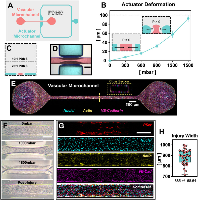
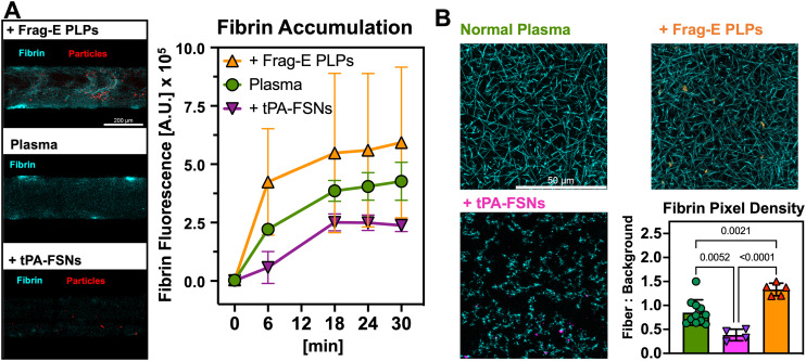
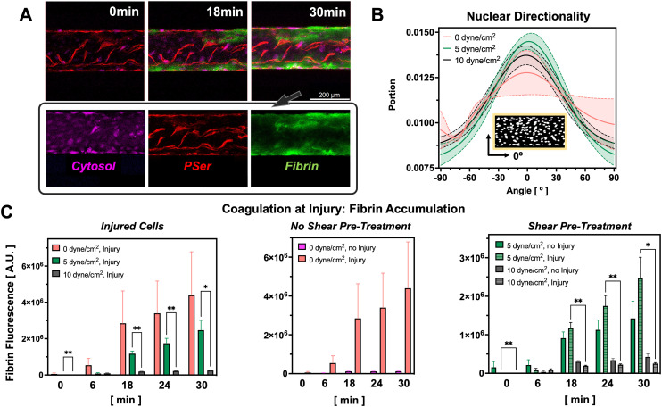
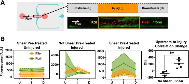
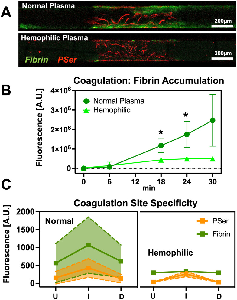
Blood coagulation is a highly regulated injury response that features polymerization of fibrin fibers to prevent the passage of blood from a damaged vascular endothelium. A growing body of research seeks to monitor coagulation in microfluidic systems but fails to capture coagulation as a response to disruption of the vascular endothelium. Here we present a device that allows compression injury of a defined segment of a microfluidic vascular endothelium and the assessment of coagulation at the injury site. This pressure injury-on-a-chip (PINCH) device allows visualization of coagulation as the accumulation of fluorescent fibrin at injury sites. Quantification of fluorescent fibrin levels upstream of and at injury sites confirm that pre-treating vascular endothelium with fluid shear stress helps capture coagulation as an injury response. We leverage the PINCH devices to demonstrate the limited coagulation response of type A hemophiliacs and evaluate the performance of hemostatic microparticles and fibrinolytic nanoparticles. Our findings and the straightforward fabrication of the PINCH devices make it a promising choice for additional screening of hemostatic therapeutics.
血液凝固是一种高度受调控的损伤反应,其特征是纤维蛋白纤维聚合,以防止血液从受损的血管内皮中流出。越来越多的研究试图在微流控系统中监测凝血过程,但未能捕捉到作为对血管内皮破坏反应的凝血过程。在此,我们展示了一种装置,该装置能够对微流控血管内皮的特定节段进行压缩损伤,并评估损伤部位的凝血情况。这种芯片上的压力损伤(PINCH)装置能够将凝血过程可视化为损伤部位荧光纤维蛋白的积累。对损伤部位上游和损伤部位荧光纤维蛋白水平的定量分析证实,用流体剪切应力对血管内皮进行预处理有助于捕捉作为损伤反应的凝血过程。我们利用PINCH装置证明了A型血友病患者有限的凝血反应,并评估了止血微粒和纤维蛋白溶解纳米颗粒的性能。我们的研究结果以及PINCH装置的简单制造使其成为进一步筛选止血治疗药物的有前景的选择。