Taube Janis M, Sunshine Joel C, Angelo Michael, Akturk Guray, Eminizer Margaret, Engle Logan L, Ferreira Cláudia S, Gnjatic Sacha, Green Benjamin, Greenbaum Shirley, Greenwald Noah F, Hedvat Cyrus V, Hollmann Travis J, Jiménez-Sánchez Daniel, Korski Konstanty, Lako Ana, Parra Edwin R, Rebelatto Marlon C, Rimm David L, Rodig Scott J, Rodriguez-Canales Jamie, Roskes Jeffrey S, Schalper Kurt A, Schenck Emanuel, Steele Keith E, Surace Michael J, Szalay Alexander S, Tetzlaff Michael T, Wistuba Ignacio I, Yearley Jennifer H, Bifulco Carlo B
Mark Foundation Center for Advanced Genomics and Imaging, Johns Hopkins University SOM, Baltimore, Maryland, USA
Bloomberg~Kimmel Institute of Cancer Immunotherapy, Johns Hopkins University SOM, Baltimore, Maryland, USA.
J Immunother Cancer. 2025 Jan 8;13(1):e008875. doi: 10.1136/jitc-2024-008875.
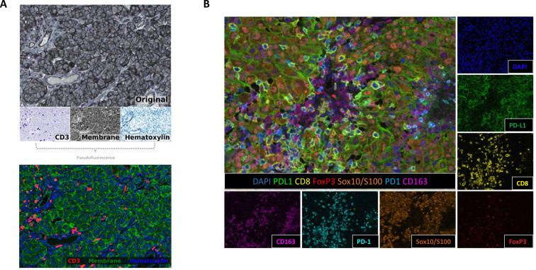
Multiplex immunohistochemistry and immunofluorescence (mIHC/IF) are emerging technologies that can be used to help define complex immunophenotypes in tissue, quantify immune cell subsets, and assess the spatial arrangement of marker expression. mIHC/IF assays require concerted efforts to optimize and validate the multiplex staining protocols prior to their application on slides. The best practice guidelines for staining and validation of mIHC/IF assays across platforms were previously published by this task force. The current effort represents a complementary manuscript for mIHC/IF analysis focused on the associated image analysis and data management.
The Society for Immunotherapy of Cancer convened a task force of pathologists and laboratory leaders from academic centers as well as experts from pharmaceutical and diagnostic companies to develop best practice guidelines for the quantitative image analysis of mIHC/IF output and data management considerations.
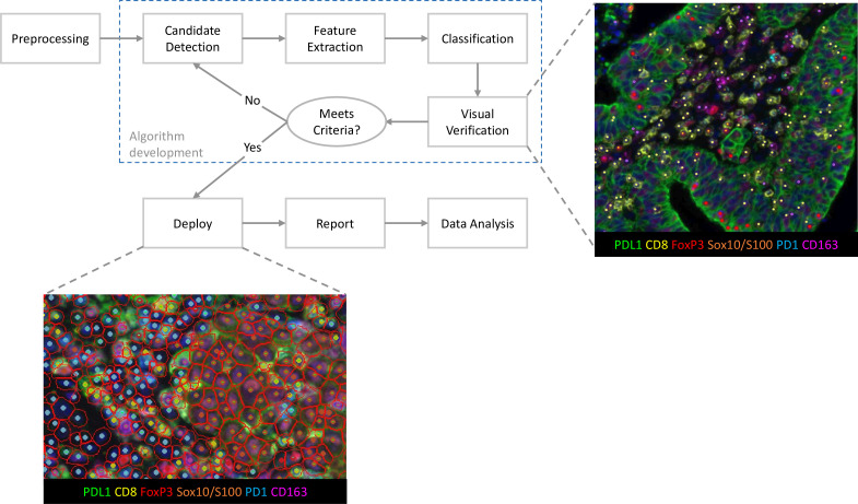
Best-practice approaches for image acquisition, color deconvolution and spectral unmixing, tissue and cell segmentation, phenotyping, and algorithm verification are reviewed. Additional quality control (QC) measures such as batch-to-batch correction and QC for assembled images are also discussed. Recommendations for sharing raw outputs, processed results, key analysis programs and source code, and representative photomicrographs from mIHC/IF assays are included. Lastly, multi-institutional harmonization efforts are described.
mIHC/IF technologies are maturing and are routinely included in research studies and moving towards clinical use. Guidelines for how to perform and standardize image analysis on mIHC/IF-stained slides will likely contribute to more comparable results across laboratories and pave the way for clinical implementation. A checklist encompassing these two-part guidelines for the generation of robust data from quantitative mIHC/IF assays will be provided in a third publication from this task force. While the current effort is mainly focused on best practices for characterizing the tumor microenvironment, these principles are broadly applicable to any mIHC/IF assay and associated image analysis.
多重免疫组化和免疫荧光(mIHC/IF)是新兴技术,可用于帮助定义组织中的复杂免疫表型、量化免疫细胞亚群以及评估标志物表达的空间排列。mIHC/IF检测在应用于玻片之前,需要协同努力来优化和验证多重染色方案。该特别工作组此前已发布了跨平台mIHC/IF检测的染色和验证最佳实践指南。当前的工作是一篇关于mIHC/IF分析的补充稿件,重点关注相关的图像分析和数据管理。
癌症免疫治疗协会召集了一个由学术中心的病理学家和实验室负责人以及制药和诊断公司的专家组成的特别工作组,以制定mIHC/IF输出定量图像分析和数据管理考量的最佳实践指南。
回顾了图像采集、颜色反卷积和光谱解混、组织和细胞分割、表型分析以及算法验证的最佳实践方法。还讨论了额外的质量控制(QC)措施,如批次间校正和组装图像的QC。包括了关于共享原始输出、处理结果、关键分析程序和源代码以及mIHC/IF检测代表性显微照片的建议。最后,描述了多机构协调工作。
mIHC/IF技术正在成熟,并常规纳入研究且正朝着临床应用发展。关于如何对mIHC/IF染色玻片进行图像分析和标准化的指南可能会有助于不同实验室获得更具可比性的结果,并为临床应用铺平道路。该特别工作组的第三篇出版物将提供一份涵盖这两部分指南的清单,用于从定量mIHC/IF检测中生成可靠数据。虽然当前的工作主要集中在表征肿瘤微环境的最佳实践上,但这些原则广泛适用于任何mIHC/IF检测及相关图像分析。