Gemechu Fitsum A, Negussie Michael A, Gebrekidan Messay, Bekele Biruk Zenebe, Mamo Elsa Wolde, Gebremariam Shimelis Nigussie
School of Medicine, College of Health Sciences, Addis Ababa University, Addis Ababa, Ethiopia.
School of Medicine, College of Health Sciences, Addis Ababa University, Addis Ababa, Ethiopia.
Int J Surg Case Rep. 2025 Feb;127:110898. doi: 10.1016/j.ijscr.2025.110898. Epub 2025 Jan 15.
Choledochal cysts are rare congenital anomalies of the bile ducts, with adult presentations being uncommon. This case is notable for its atypical presentation in a young adult, mimicking a hydatid cyst in a region where echinococcosis is endemic.
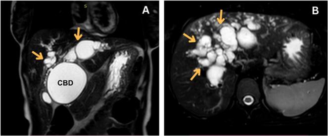
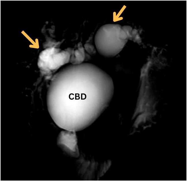
A 22-year-old female presented with a 3-month history of progressive jaundice, accompanied by 5 months of epigastric and right upper quadrant pain, dark urine, pale stools, pruritus, and significant weight loss. She reported a prior admission for cholangitis, treated with antibiotics. Examination revealed stable vital signs, icteric sclerae, right upper quadrant tenderness, and scratch marks on the skin. Laboratory investigations showed elevated liver enzymes and hyperbilirubinemia (total bilirubin = 26 mg/dL, direct bilirubin = 20.5 mg/dL). Initial imaging studies, including ultrasound and CT, suggested a hydatid cyst of the liver. However, MRCP revealed dilated intrahepatic and extrahepatic bile ducts, consistent with a Type IV-A choledochal cyst. The patient underwent cholecystectomy, extrahepatic bile duct excision, and Roux-en-Y cysto-jejunostomy. Histopathological analysis confirmed the diagnosis without evidence of malignancy. She recovered uneventfully, with no complications reported during a 6-month follow-up.
This case highlights the diagnostic challenges in differentiating choledochal cysts from hydatid cysts, particularly in endemic regions. The use of MRCP was pivotal in achieving an accurate diagnosis and guiding definitive management. Early surgical intervention minimized the risks of complications and malignancy.
Type IV-A choledochal cysts can present atypically, mimicking hydatid cysts. Advanced imaging, especially MRCP, is critical for accurate diagnosis and management.
胆管囊肿是一种罕见的胆管先天性异常,成人病例并不常见。该病例的显著特点是在一名年轻成年人中出现非典型表现,在棘球蚴病流行地区类似包虫囊肿。
一名22岁女性,有3个月进行性黄疸病史,伴有5个月上腹部和右上腹疼痛、深色尿液、浅色粪便、瘙痒和显著体重减轻。她曾因胆管炎入院,接受过抗生素治疗。检查发现生命体征稳定,巩膜黄染,右上腹压痛,皮肤有抓痕。实验室检查显示肝酶升高和高胆红素血症(总胆红素 = 26 mg/dL,直接胆红素 = 20.5 mg/dL)。包括超声和CT在内的初始影像学检查提示肝脏有包虫囊肿。然而,磁共振胰胆管造影(MRCP)显示肝内和肝外胆管扩张,符合IV-A型胆管囊肿。患者接受了胆囊切除术、肝外胆管切除术和Roux-en-Y囊肿空肠吻合术。组织病理学分析确诊,未发现恶性证据。她恢复顺利,6个月随访期间未报告并发症。
该病例突出了在区分胆管囊肿和包虫囊肿时面临的诊断挑战,特别是在流行地区。MRCP的使用对于准确诊断和指导最终治疗至关重要。早期手术干预将并发症和恶性肿瘤的风险降至最低。
IV-A型胆管囊肿可表现为非典型,类似包虫囊肿。先进的影像学检查,尤其是MRCP,对于准确诊断和治疗至关重要。