Zhao Hanqiu, Ma Xiaokuang, Chen Peng, Liu Bin, Wei Jing, Zhang John, Desai Ankit A, Frump Andrea L, Rafikova Olga, Fallon Michael B, Qiu Shenfeng, Dai Zhiyu
Department of Internal Medicine, University of Arizona College of Medicine-Phoenix, Phoenix, AZ 85004, USA.
Division of Pulmonary, Critical Care Medicine, John T. Milliken Department of Medicine, Washington University School of Medicine in Saint Louis, Saint Louis, MO 63110, USA.
J Respir Biol Transl Med. 2025;2(2). doi: 10.70322/jrbtm.2025.10004. Epub 2025 May 15.
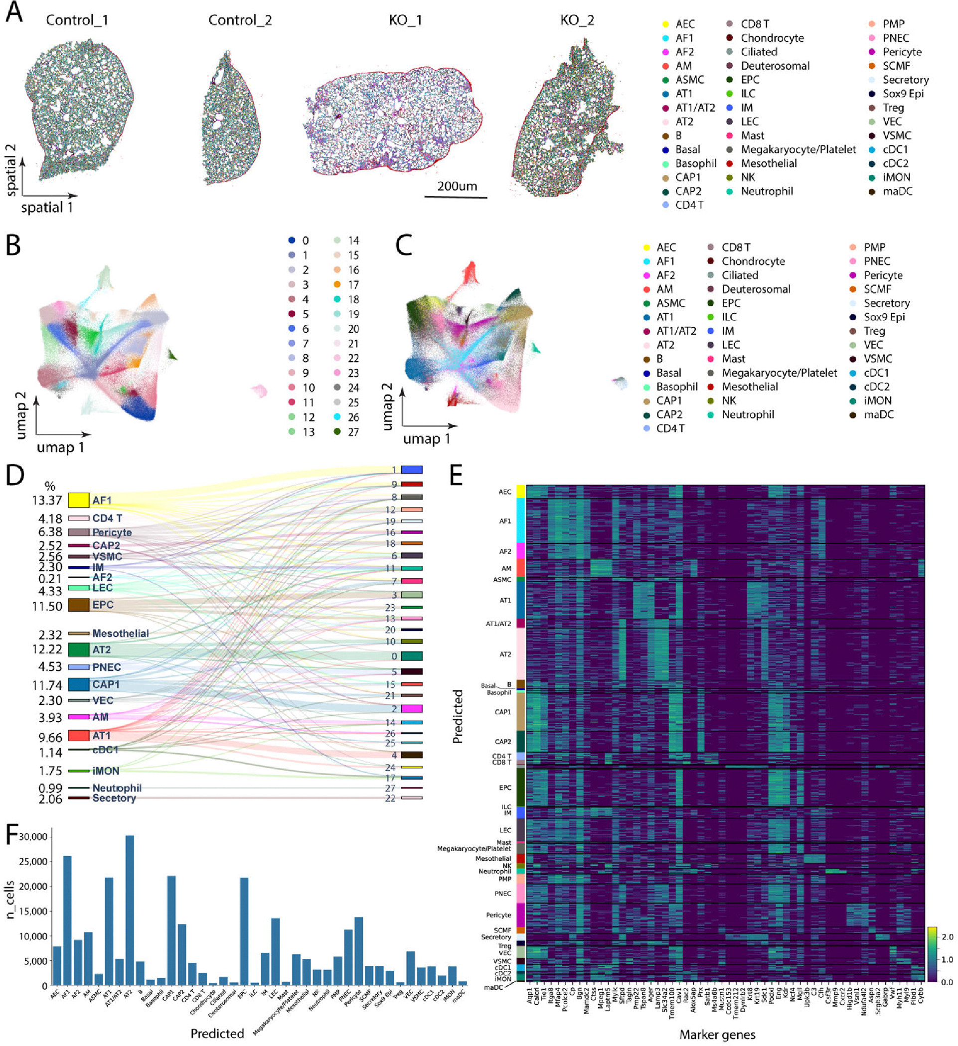
Spatial transcriptomics technologies have emerged as powerful tools for understanding cellular identity and function within the natural spatial context of tissues. Traditional transcriptomics techniques, such as bulk and single-cell RNA sequencing, lose this spatial information, which is critical for addressing many biological questions. Here, we present a protocol for high-resolution spatial transcriptomics using fixed frozen mouse lung sections mounted on 10X Genomics Xenium slides. This method integrates multiplexed fluorescent in situ hybridization (FISH) with high-throughput imaging to reveal the spatial distribution of mRNA molecules in lung tissue sections, allowing detailed analysis of gene expression changes in a mouse model of pulmonary hypertension (PH). We compared two tissue preparation methods, fixed frozen and fresh frozen, for compatibility with the Xenium platform. Our fixed frozen approach, utilizing a free-floating technique to mount thin lung sections onto Xenium slides at room temperature, preserved tissue integrity and maximized the imaging area, resulting in high-fidelity spatial transcriptomics data. Using a predesigned 379-gene mouse panel, we identified 40 major lung cell types. We detected key cellular changes in PH, including an increase in arterial endothelial cells (AECs) and fibroblasts, alongside a reduction in capillary endothelial cells (CAP1 and CAP2). Through differential gene expression analysis, we observed markers of endothelial-to-mesenchymal transition and fibroblast activation in PH lungs. High-resolution spatial mapping further confirmed increased arterialization in the distal microvasculature. These findings underscore the utility of spatial transcriptomics in preserving the native tissue architecture and enhancing our understanding of cellular heterogeneity in disease. Our protocol provides a reliable method for integrating spatial and transcriptomic data using fixed frozen lung tissues, offering significant potential for future studies in complex diseases such as PH.
空间转录组学技术已成为在组织的自然空间背景下理解细胞身份和功能的强大工具。传统的转录组学技术,如批量和单细胞RNA测序,会丢失这种空间信息,而这种信息对于解决许多生物学问题至关重要。在这里,我们展示了一种使用安装在10X Genomics Xenium载玻片上的固定冷冻小鼠肺切片进行高分辨率空间转录组学的方案。该方法将多重荧光原位杂交(FISH)与高通量成像相结合,以揭示肺组织切片中mRNA分子的空间分布,从而能够详细分析肺动脉高压(PH)小鼠模型中的基因表达变化。我们比较了两种组织制备方法,即固定冷冻和新鲜冷冻,以评估它们与Xenium平台的兼容性。我们的固定冷冻方法采用自由漂浮技术在室温下将薄肺切片安装到Xenium载玻片上,保持了组织完整性并最大化了成像面积,从而获得了高保真的空间转录组学数据。使用预先设计的379个基因的小鼠基因panel,我们鉴定出40种主要的肺细胞类型。我们检测到PH中的关键细胞变化,包括动脉内皮细胞(AEC)和成纤维细胞的增加,同时毛细血管内皮细胞(CAP1和CAP2)减少。通过差异基因表达分析,我们在PH肺中观察到内皮-间充质转化和成纤维细胞激活的标志物。高分辨率空间映射进一步证实了远端微血管中动脉化增加。这些发现强调了空间转录组学在保留天然组织结构和增强我们对疾病中细胞异质性理解方面的实用性。我们的方案提供了一种使用固定冷冻肺组织整合空间和转录组数据的可靠方法,为未来在诸如PH等复杂疾病的研究中提供了巨大潜力。