• 文献检索
  • 文档翻译
  • 深度研究
  • 学术资讯
  • Suppr Zotero 插件Zotero 插件
  • 邀请有礼
  • 套餐&价格
  • 历史记录
应用&插件
Suppr Zotero 插件Zotero 插件浏览器插件Mac 客户端Windows 客户端微信小程序
定价
高级版会员购买积分包购买API积分包
服务
文献检索文档翻译深度研究API 文档MCP 服务
关于我们
关于 Suppr公司介绍联系我们用户协议隐私条款
关注我们

Suppr 超能文献

核心技术专利:CN118964589B侵权必究
粤ICP备2023148730 号-1Suppr @ 2026

文献检索

告别复杂PubMed语法,用中文像聊天一样搜索,搜遍4000万医学文献。AI智能推荐,让科研检索更轻松。

立即免费搜索

文件翻译

保留排版,准确专业,支持PDF/Word/PPT等文件格式,支持 12+语言互译。

免费翻译文档

深度研究

AI帮你快速写综述,25分钟生成高质量综述,智能提取关键信息,辅助科研写作。

立即免费体验

成人主动脉瓣狭窄。通过连续波多普勒超声心动图对压力差进行无创估计。

Aortic stenosis in adults. Non-invasive estimation of pressure differences by continuous wave Doppler echocardiography.

作者信息

Hegrenaes L, Hatle L

出版信息

Br Heart J. 1985 Oct;54(4):396-404. doi: 10.1136/hrt.54.4.396.

DOI:10.1136/hrt.54.4.396
PMID:4052281
原文链接:https://pmc.ncbi.nlm.nih.gov/articles/PMC481918/
Abstract

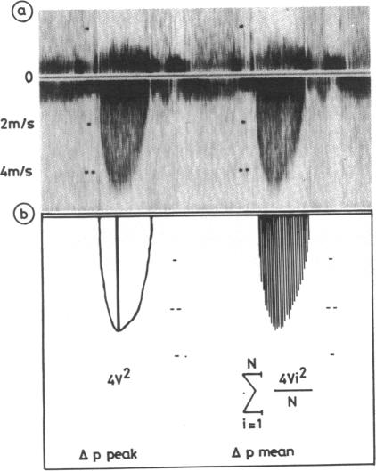
The peak and mean aortic transvalvar pressure differences measured invasively and non-invasively by continuous wave Doppler echocardiography were compared in 87 consecutive patients with aortic stenosis. The mean values were calculated from the maximal velocities of the aortic jet recorded with a spectral display of the Doppler frequency shifts and by applying a modified Bernoulli equation. Technically satisfactory velocity curves for estimating the mean pressure differences could not be obtained in three patients and invasive measurements were not obtained in two. In all patients the peak transvalvar pressure difference was calculated since the aortic jet was identified non-invasively. The peak and mean pressure differences measured invasively and non-invasively correlated well--with only minor underestimation of the pressure differences measured with the Doppler technique--regardless of age, sex, and the presence or absence of aortic valvar regurgitation, or other valvar lesions. With a systematic search for the highest velocities in the aortic jet and with on line spectral analysis of the Doppler frequencies the peak and the mean aortic pressure differences can be determined non-invasively with a high degree of precision in almost all patients.

摘要

采用连续波多普勒超声心动图对87例连续性主动脉瓣狭窄患者进行有创和无创测量,比较主动脉跨瓣压差的峰值和平均值。平均值通过多普勒频移频谱显示记录的主动脉射流最大速度并应用改良的伯努利方程计算得出。3例患者未能获得技术上满意的用于估计平均压差的速度曲线,2例未进行有创测量。在所有患者中,由于通过无创方法识别了主动脉射流,因此计算了跨瓣压差峰值。无论年龄、性别、有无主动脉瓣反流或其他瓣膜病变,有创和无创测量的峰值和平均压差相关性良好,仅多普勒技术测量的压差略有低估。通过系统寻找主动脉射流中的最高速度并对多普勒频率进行在线频谱分析,几乎所有患者都能以高度的精确性无创测定主动脉压差的峰值和平均值。

https://cdn.ncbi.nlm.nih.gov/pmc/blobs/a01b/481918/b19fffb8f085/brheartj00118-0058-a.jpg
https://cdn.ncbi.nlm.nih.gov/pmc/blobs/a01b/481918/0a7888cc85fa/brheartj00118-0057-a.jpg
https://cdn.ncbi.nlm.nih.gov/pmc/blobs/a01b/481918/b19fffb8f085/brheartj00118-0058-a.jpg
https://cdn.ncbi.nlm.nih.gov/pmc/blobs/a01b/481918/0a7888cc85fa/brheartj00118-0057-a.jpg
https://cdn.ncbi.nlm.nih.gov/pmc/blobs/a01b/481918/b19fffb8f085/brheartj00118-0058-a.jpg

相似文献

1
Aortic stenosis in adults. Non-invasive estimation of pressure differences by continuous wave Doppler echocardiography.成人主动脉瓣狭窄。通过连续波多普勒超声心动图对压力差进行无创估计。
Br Heart J. 1985 Oct;54(4):396-404. doi: 10.1136/hrt.54.4.396.
2
Evaluation of aortic stenosis severity: role of contrast echocardiography in comparison with conventional echocardiography and cardiac catheterization.主动脉瓣狭窄严重程度的评估:对比超声心动图与传统超声心动图及心导管检查相比的作用。
Rev Port Cardiol. 2002 May;21(5):555-72.
3
[Continuous wave Doppler echocardiographic assessment of aortic stenosis and aortic regurgitation].[主动脉瓣狭窄和主动脉瓣反流的连续波多普勒超声心动图评估]
J Cardiogr. 1985 Mar;15(1):111-21.
4
Quantification of transvalvular pressure differences in aortic stenosis by Doppler ultrasound.通过多普勒超声对主动脉瓣狭窄跨瓣压差进行定量分析。
Int J Cardiol. 1985 Feb;7(2):121-8. doi: 10.1016/0167-5273(85)90350-x.
5
Determination of the mean pressure gradient in aortic stenosis by Doppler echocardiography.通过多普勒超声心动图测定主动脉瓣狭窄时的平均压力阶差。
Eur Heart J. 1985 Dec;6(12):999-1005. doi: 10.1093/oxfordjournals.eurheartj.a061821.
6
[Severity of aortic stenosis assessed by Doppler techniques].[通过多普勒技术评估主动脉瓣狭窄的严重程度]
J Cardiol. 1987 Dec;17(4):807-16.
7
Limitations in assessing the severity of aortic stenosis by Doppler gradients.通过多普勒梯度评估主动脉瓣狭窄严重程度的局限性。
Br Heart J. 1988 May;59(5):551-5. doi: 10.1136/hrt.59.5.551.
8
[Quantitative evaluation of aortic stenosis using continuous wave Doppler].[使用连续波多普勒对主动脉瓣狭窄进行定量评估]
Zhonghua Xin Xue Guan Bing Za Zhi. 1989 Feb;17(1):7-9, 61.
9
Comparison of high pulse repetition frequency and continuous wave Doppler echocardiography in the assessment of high flow velocity in patients with valvular stenosis and regurgitation.
J Am Coll Cardiol. 1985 Sep;6(3):565-71. doi: 10.1016/s0735-1097(85)80114-5.
10
Aortic valve area discrepancy by Gorlin equation and Doppler echocardiography continuity equation: relationship to flow in patients with valvular aortic stenosis.根据 Gorlin 公式和多普勒超声心动图连续性方程得出的主动脉瓣面积差异:与瓣膜性主动脉狭窄患者血流的关系
Can J Cardiol. 2000 Aug;16(8):985-92.

引用本文的文献

1
Non-invasive myocardial work in aortic stenosis: validation and improvement in left ventricular pressure estimation.主动脉瓣狭窄患者的无创心肌做功:左心室压力估计的验证和改进。
Eur Heart J Cardiovasc Imaging. 2024 Jan 29;25(2):201-212. doi: 10.1093/ehjci/jead227.
2
Relative pressure estimation from 4D flow MRI using generalized Bernoulli equation in a phantom model of arterial stenosis.基于广义伯努利方程的 4D 血流 MRI 在动脉狭窄仿体模型中相对压力的估计。
MAGMA. 2022 Oct;35(5):733-748. doi: 10.1007/s10334-022-01001-x. Epub 2022 Feb 17.
3
Towards improving the accuracy of aortic transvalvular pressure gradients: rethinking Bernoulli.

本文引用的文献

1
Non-invasive assessment of aortic stenosis by Doppler ultrasound.通过多普勒超声对主动脉瓣狭窄进行无创评估。
Br Heart J. 1980 Mar;43(3):284-92. doi: 10.1136/hrt.43.3.284.
2
Noninvasive assessment and differentiation of left ventricular outflow obstruction with Doppler ultrasound.应用多普勒超声对左心室流出道梗阻进行无创评估与鉴别
Circulation. 1981 Aug;64(2):381-7. doi: 10.1161/01.cir.64.2.381.
3
Quantification of pressure gradients across stenotic valves by Doppler ultrasound.通过多普勒超声对狭窄瓣膜两端的压力阶差进行定量分析。
为了提高主动脉瓣跨瓣压差的准确性:重新思考伯努利。
Med Biol Eng Comput. 2020 Aug;58(8):1667-1679. doi: 10.1007/s11517-020-02186-w. Epub 2020 May 26.
4
Non-Invasive Assessment of Intravascular Pressure Gradients: A Review of Current and Proposed Novel Methods.血管内压力梯度的非侵入性评估:当前及新型方法综述
Diagnostics (Basel). 2018 Dec 29;9(1):5. doi: 10.3390/diagnostics9010005.
5
MRI for diagnosing aortic valve stenosis: a comparison study of MRI and ultrasound.磁共振成像用于诊断主动脉瓣狭窄:磁共振成像与超声检查的对比研究
Neth Heart J. 2005 Nov;13(11):387-392.
6
High transvalvular pressure gradients on intraoperative transesophageal echocardiography after aortic valve replacement: what does it mean?主动脉瓣置换术后术中经食管超声心动图显示的高跨瓣压差:这意味着什么?
HSR Proc Intensive Care Cardiovasc Anesth. 2009;1(4):7-18.
7
[Perioperative echocardiography: basic principles].[围手术期超声心动图:基本原则]
Anaesthesist. 2006 Mar;55(3):337-61. doi: 10.1007/s00101-006-0992-9.
8
Discrepancy between Doppler and catheter measurements of pressure gradients across small-size prosthetic valve.
Jpn J Thorac Cardiovasc Surg. 2005 Feb;53(2):64-8. doi: 10.1007/s11748-005-0002-1.
9
Developments in cardiovascular ultrasound. Part 3: Cardiac applications.心血管超声的发展。第3部分:心脏应用。
Med Biol Eng Comput. 1998 Sep;36(5):529-43. doi: 10.1007/BF02524420.
10
Planimetry of aortic valve area using multiplane transoesophageal echocardiography is not a reliable method for assessing severity of aortic stenosis.使用多平面经食管超声心动图测量主动脉瓣面积并非评估主动脉瓣狭窄严重程度的可靠方法。
Heart. 1997 Jul;78(1):68-73. doi: 10.1136/hrt.78.1.68.
J Am Coll Cardiol. 1983 Oct;2(4):707-18. doi: 10.1016/s0735-1097(83)80311-8.
4
Noninvasive prediction of transvalvular pressure gradient in patients with pulmonary stenosis by quantitative two-dimensional echocardiographic Doppler studies.通过定量二维超声心动图多普勒研究对肺动脉狭窄患者跨瓣压差进行无创预测。
Circulation. 1983 Apr;67(4):866-71. doi: 10.1161/01.cir.67.4.866.
5
In vitro verification of Doppler prediction of transvalve pressure gradient and orifice area in stenosis.狭窄时经瓣压力阶差和瓣口面积的多普勒预测的体外验证
Am J Cardiol. 1984 May 1;53(9):1369-73. doi: 10.1016/0002-9149(84)90096-1.
6
Noninvasive determination of pressure gradients in children: two methods employing pulsed Doppler echocardiography.
J Am Coll Cardiol. 1984 Jan;3(1):179-92. doi: 10.1016/s0735-1097(84)80446-5.
7
Evaluation of aortic stenosis by continuous wave Doppler ultrasound.
J Am Coll Cardiol. 1984 Jan;3(1):150-6. doi: 10.1016/s0735-1097(84)80442-8.
8
Prediction of the severity of left ventricular outflow tract obstruction by quantitative two-dimensional echocardiographic Doppler studies.通过定量二维超声心动图多普勒研究预测左心室流出道梗阻的严重程度。
Circulation. 1983 Aug;68(2):348-54. doi: 10.1161/01.cir.68.2.348.
9
Evaluation of systolic murmurs by Doppler ultrasonography.通过多普勒超声心动图评估收缩期杂音。
Br Heart J. 1983 Oct;50(4):337-42. doi: 10.1136/hrt.50.4.337.
10
Determination of pressure gradient in mitral stenosis with a non-invasive ultrasound Doppler technique.
Acta Med Scand. 1976;199(6):455-60. doi: 10.1111/j.0954-6820.1976.tb06763.x.