Itoh Kosuke, Konoike Naho, Iwaoki Haruhiko, Igarashi Hironaka, Nakamura Katsuki
Center for Integrated Human Brain Science, Brain Research Institute, Niigata University, Niigata, Japan.
Cognitive Neuroscience Section, Primate Research Institute, Kyoto University, Inuyama, Aichi, 951-8585, Japan.
Neuroimage Rep. 2022 Jun 25;2(3):100116. doi: 10.1016/j.ynirp.2022.100116. eCollection 2022 Sep.
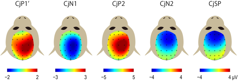
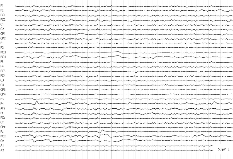
Noninvasive electroencephalogram (EEG) in unanesthetized nonhuman primates is useful for basic science and biomedical applications but presents several technical challenges. In particular, multiple EEG electrode application to the scalp of small-sized monkeys, such as common marmosets, has been difficult, if not impossible. Here, we describe a novel approach, using the "dip-in electrode" method, which enables multichannel noninvasive EEG recording in a marmoset through short and small silicone tubes, with an internal diameter of 4 mm. First, the cut face of the tube was glued to the shaved scalp. Then, the tube was filled with the electrode gel, and a small electrode (4 mm in diameter) was dipped into the gel to contact the skin electrically while being electrically isolated from the neighboring channels. The minimum inter-electrode distance was 6 mm, which was limited by the outer diameter of the tubes. A proof of concept was provided through successful 32-channel recording of scalp EEG and topographic mapping of the cortical auditory evoked potentials in an unanesthetized marmoset. This had not been possible with the existing methods using gauze or tape for electrode fixation. Our method provides a feasible means for noninvasive scalp recording of EEG and evoked potentials in marmoset monkeys and possibly other nonhuman primates and non-primate mammals.
在未麻醉的非人灵长类动物中进行无创脑电图(EEG)检测,对基础科学和生物医学应用很有用,但存在一些技术挑战。特别是,将多个EEG电极应用于小型猴子(如普通狨猴)的头皮一直很困难,甚至可以说是不可能的。在此,我们描述了一种新颖的方法,即使用“浸入式电极”方法,通过内径为4毫米的短而小的硅胶管,在狨猴中实现多通道无创EEG记录。首先,将管子的切割面粘在剃毛的头皮上。然后,在管中填充电极凝胶,将一个小电极(直径4毫米)浸入凝胶中,使其与皮肤电接触,同时与相邻通道电隔离。电极间的最小距离为6毫米,这受到管子外径的限制。通过在未麻醉的狨猴中成功进行32通道头皮EEG记录和皮质听觉诱发电位的地形图绘制,提供了概念验证。使用纱布或胶带固定电极的现有方法无法做到这一点。我们的方法为在狨猴以及可能的其他非人灵长类动物和非灵长类哺乳动物中进行EEG和诱发电位的无创头皮记录提供了一种可行的手段。