Greaves M F, Roitt I M
Clin Exp Immunol. 1968 Jun;3(5):393-412.
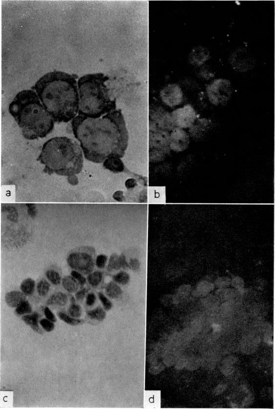
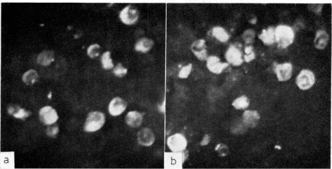
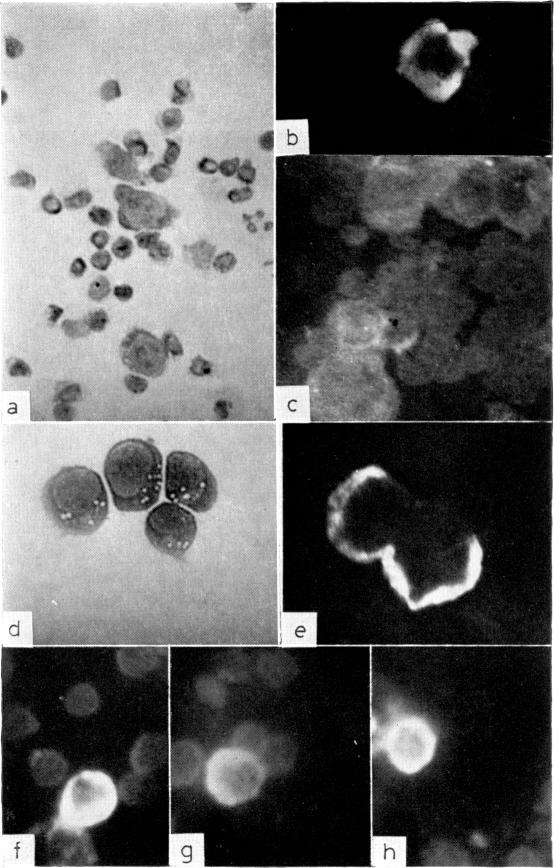
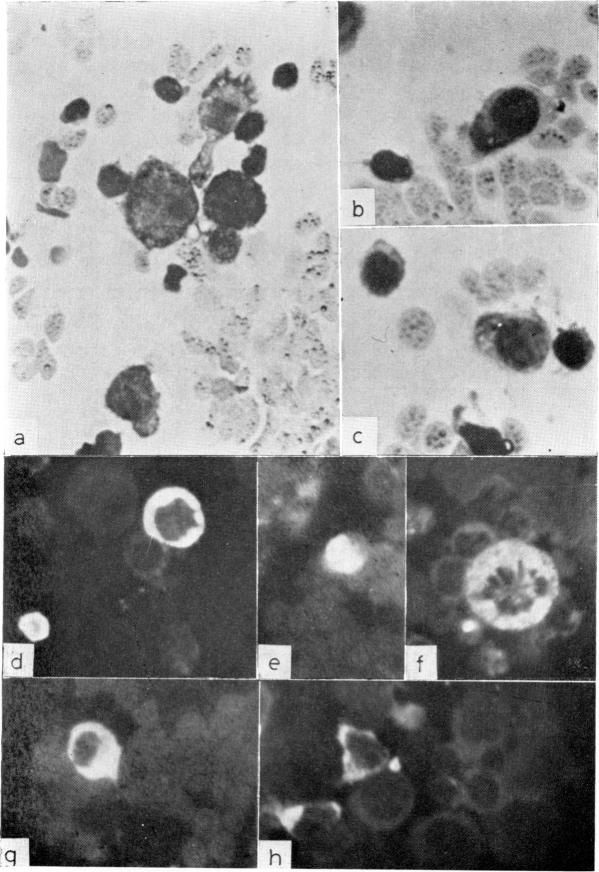
Amino acid incorporation techniques and immunofluorescence have been used to investigate the effect of mitogenic substances on immunoglobulin synthesis by human peripheral blood lymphocytes . Radioelectrophoresis, radioimmunoelectrophoresis and controlled immunological precipitation methods suggest that only a small amount of immunoglobulin is synthesized in the culture system used. Immunofluorescent staining of fixed cell preparations showed that during the first 24 hr in culture only a small percentage of cells reacted positively for immunoglobulin; after 24 hr these cells were no longer demonstrable. This suggests that the small amount of immunoglobulin detected was synthesized during the first few hours in culture by these cells, having the morphological appearance of medium lymphocytes. The slight enhancement of immunoglobulin synthesis obtained in one experiment with phytohaemagglutinin (PHA) probably occurred within this same cell type since after 24 hr no cells in the transformed cultures could be stained by the fluorescent anti-immunoglobulin. Fixed preparations of blast cells obtained by stimulation with anti-lymphocytic serum and staphylococcal filtrate also gave negative reactions. However, using a staining technique with suspensions of viable cells, it was possible to demonstrate positive staining for immunoglobulins with PHA stimulated cells as previously described by Ripps & Hirschhorn (1967). A number of controls suggest that this reaction depends upon the presence of exposed immunoglobulin groups or markers on the cell surface and that intracytoplasmic staining is the result of endocytosis of conjugate. In contrast with the negative results obtained with PHA-transformed blasts, a small percentage of lymphocytes from cultures stimulated by pokeweed or tuberculin reacted positively when fixed preparations were stained with conjugated anti-immunoglobulin.
氨基酸掺入技术和免疫荧光法已被用于研究促有丝分裂物质对人外周血淋巴细胞免疫球蛋白合成的影响。放射电泳、放射免疫电泳和可控免疫沉淀法表明,在所使用的培养系统中仅合成了少量免疫球蛋白。固定细胞制剂的免疫荧光染色显示,在培养的最初24小时内,只有一小部分细胞对免疫球蛋白呈阳性反应;24小时后,这些细胞不再可见。这表明检测到的少量免疫球蛋白是在培养的最初几个小时内由这些具有中等淋巴细胞形态外观的细胞合成的。在一项用植物血凝素(PHA)进行的实验中获得的免疫球蛋白合成的轻微增强可能发生在同一细胞类型中,因为在24小时后,转化培养物中的细胞均不能被荧光抗免疫球蛋白染色。用抗淋巴细胞血清和葡萄球菌滤液刺激获得的母细胞固定制剂也呈阴性反应。然而,使用活细胞悬液的染色技术,可以如Ripps和Hirschhorn(1967年)先前所述,证明PHA刺激的细胞对免疫球蛋白呈阳性染色。许多对照表明,这种反应取决于细胞表面暴露的免疫球蛋白基团或标志物的存在,而胞质内染色是结合物内吞作用的结果。与PHA转化的母细胞获得的阴性结果相反,当用结合的抗免疫球蛋白对固定制剂进行染色时,来自商陆或结核菌素刺激培养物的一小部分淋巴细胞呈阳性反应。