Morgan C, Rose H M, Mednis B
J Virol. 1968 May;2(5):507-16. doi: 10.1128/JVI.2.5.507-516.1968.
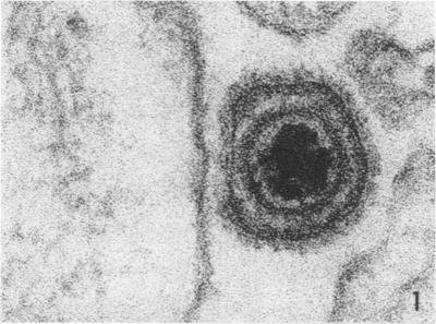
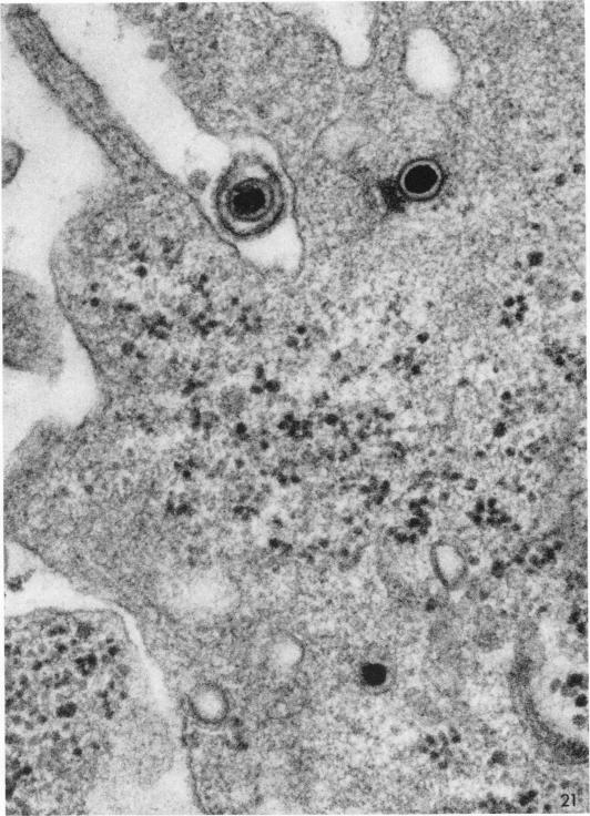
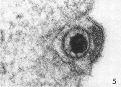
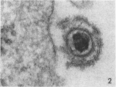
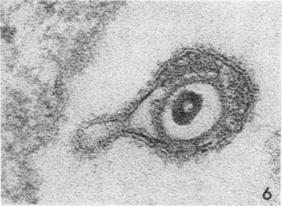
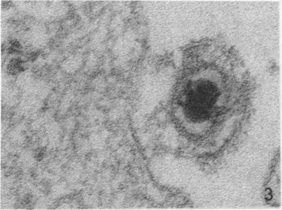
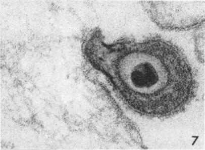
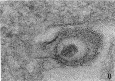
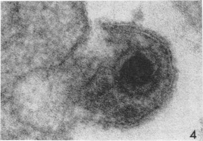
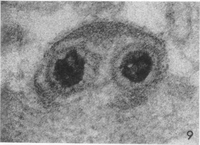
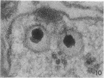
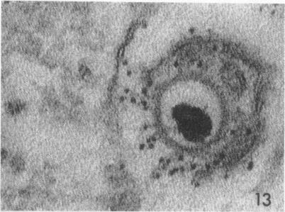
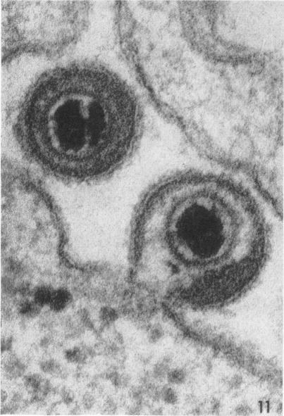
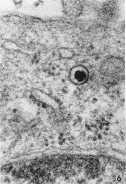
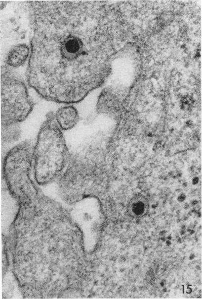
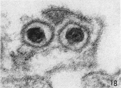
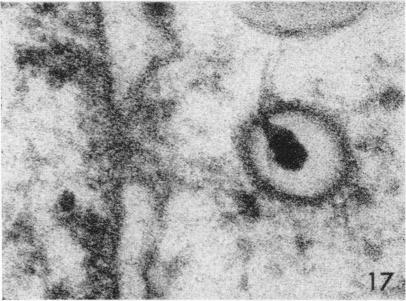
Although capsids of herpes simplex virus were encountered within phagocytic vesicles, they were more commonly observed free within the cytoplasm. Stages in the release of virus from vesicles were not seen. There appeared to be five distinct steps in the process whereby the virus initiates infection: attachment, digestion of the viral envelope, digestion of the cell wall, passage of the capsid directly into the cytoplasm, and digestion of the capsid with release of the core. Antibody probably interferes with the first two stages.
尽管在吞噬小泡内发现了单纯疱疹病毒的衣壳,但它们更常见于细胞质中游离存在。未观察到病毒从囊泡中释放的阶段。病毒引发感染的过程似乎有五个不同的步骤:附着、病毒包膜的消化、细胞壁的消化、衣壳直接进入细胞质以及衣壳消化并释放核心。抗体可能会干扰前两个阶段。