Black M M, Epstein W L
Am J Pathol. 1974 Feb;74(2):263-74.
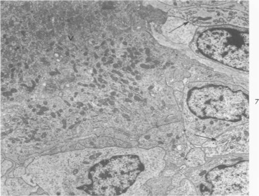
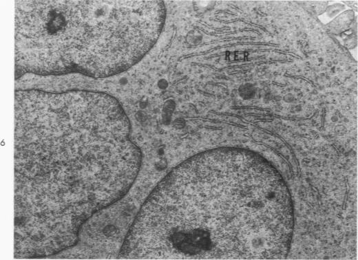
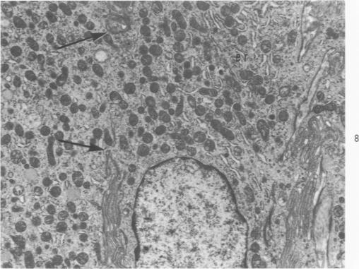
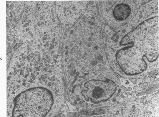
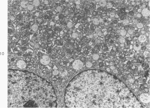
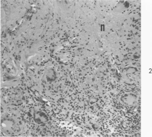
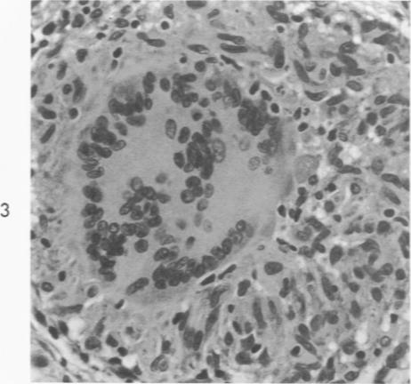
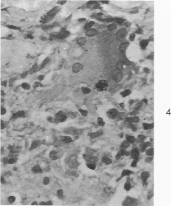
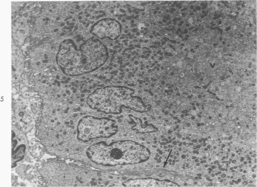
Organized epithelioid cell granulomas were produced experimentally by injecting intradermally dilute suspensions of zirconium and beryllium salts into individuals who had been previously sensitized to these metals. Biopsies were obtained at intervals of between 5 days and 13 months later, and the specimens were fixed and prepared for light and electron microscopy. Tritiated thymidine was injected into a number of the granulomas; the biopsy specimens were secured from 40 minutes to 2 or more weeks later, and the tissues were processed for light microscopic autoradiography. Giant cells occurred commonly, both within organized tubercles and in relation to areas of necrosis, and had markedly different cytoplasmic features from typical secretory epithelioid cells which enabled them to be readily recognized at scanning magnifications. The characteristic hallmark of these giant cells was the presence of myriads of small mitochondria adjacent to nuclei with numerous membrane-lined vesicles in the center of the cell. Giant cells occurred mainly in edematous disrupted tubercles. In these tubercles, epithelioid cells contained cytoplasmic components more like giant cells. Direct evidence of cell fusion was not seen, although fusion of membranes seemed to occur between cells having similar cytoplasmic features. The failure to find labeled nuclei in any giant cells 40 minutes after injection of tritiated thymidine indicates that normal nuclear division does not occur within giant cells. We postulate that epithelioid cells containing vesicles develop in damaged and necrotic areas, and that mainly this type of epithelioid cell fuses to form giant cells.
通过向先前已对锆盐和铍盐致敏的个体皮内注射稀释的锆盐和铍盐悬浮液,实验性地产生了有组织的上皮样细胞肉芽肿。在5天至13个月的间隔时间获取活检标本,将标本固定并制备用于光镜和电镜检查。向一些肉芽肿中注射氚标记的胸腺嘧啶核苷;在注射后40分钟至2周或更长时间获取活检标本,并对组织进行光镜放射自显影处理。巨细胞常见于有组织的结节内以及与坏死区域相关的部位,其细胞质特征与典型的分泌性上皮样细胞明显不同,这使得它们在扫描放大倍数下易于识别。这些巨细胞的特征性标志是在细胞核附近存在无数小线粒体,细胞中央有许多膜包被的小泡。巨细胞主要出现在水肿性破坏的结节中。在这些结节中,上皮样细胞含有更类似于巨细胞的细胞质成分。尽管在具有相似细胞质特征的细胞之间似乎发生了膜融合,但未见到细胞融合的直接证据。在注射氚标记的胸腺嘧啶核苷40分钟后,在任何巨细胞中均未发现标记的细胞核,这表明巨细胞内不会发生正常的核分裂。我们推测,含有小泡的上皮样细胞在受损和坏死区域形成,并且主要是这种类型的上皮样细胞融合形成巨细胞。