Whittlestone P, Lemcke R M, Olds R J
J Hyg (Lond). 1972 Sep;70(3):387-407. doi: 10.1017/s0022172400062975.
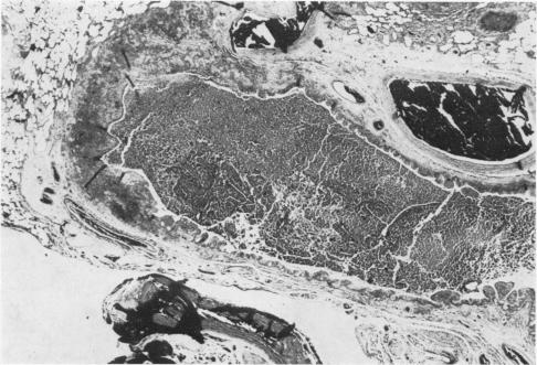
Mycoplasma pulmonis was isolated from the pneumonic lung of a rat. Two groups of mycoplasma-free rats were inoculated, one with a culture of the M. pulmonis strain which had been cloned four times (group A) and the other with a lung homogenate of the rat from which the strain had been isolated (group B). A third group (C) consisted of uninoculated control animals. Each group was kept in strict isolation and allowed to breed so that the progeny was naturally exposed to any pathogens present in the inoculated animals. After different periods of exposure, rats were autopsied, respiratory tracts and inner ears were cultured for mycoplasmas and bacteria, and sera were tested for complement-fixing antibodies to murine mycoplasmas.In group-A rats, M. pulmonis was consistently isolated from the inner ears or lungs from 50 to 715 days after exposure. Complement-fixing antibody to M. pulmonis was detected 20 days after inoculation, but in the naturally exposed progeny antibody took longer than 50 days to develop. Antibodies to the other known mycoplasmas of murine origin, M. arthritidis and M. neurolyticum, were never found. Purulent otitis interna was consistently found from day 55 onwards, while lung lesions were first observed at 85 days and persisted to 715 days. Pulmonary lesions developed more slowly in inoculated parents than in exposed progeny. Similar results were found in group-B rats, which were examined up to 441 days after inoculation. Uninoculated group-C rats were examined up to 768 days of age, but M. pulmonis was not recovered; of the 54 animals whose serum was tested all were negative to the three species of mycoplasmas, except one which had a titre of 16 with M. pulmonis. Pneumonia, bronchiectasis or lymphoreticular hyperplasia were not seen in any of these control rats. Bacterial respiratory pathogens were not isolated from rats in any of the groups, nor was antibody to Sendai virus detected.The results suggest that M. pulmonis alone can cause pneumonia and bronchiectasis in rats since mechanical carry-over of another pathogen with the initial cloned inoculum is very unlikely and there was no evidence for the participation of any other rat pathogen. The respiratory disease induced by the cloned culture was comparable with that induced by the lung homogenate, and with the well-known syndrome of chronic respiratory disease and bronchiectasis in the rat.
从一只患肺炎大鼠的肺中分离出了肺支原体。将两组无支原体大鼠进行接种,一组接种已克隆4次的肺支原体菌株培养物(A组),另一组接种分离出该菌株的大鼠的肺匀浆(B组)。第三组(C组)由未接种的对照动物组成。每组严格隔离饲养并使其繁殖,以便后代自然接触接种动物体内存在的任何病原体。在不同的暴露期后,对大鼠进行解剖,对呼吸道和内耳进行支原体和细菌培养,并检测血清中针对鼠支原体的补体结合抗体。在A组大鼠中,暴露后50至715天,始终能从内耳或肺中分离出肺支原体。接种后20天检测到针对肺支原体的补体结合抗体,但在自然暴露的后代中,抗体的产生时间超过50天。从未发现针对鼠源其他已知支原体,即关节炎支原体和溶神经支原体的抗体。从第55天起始终能发现化脓性内耳炎,而肺部病变最早在85天观察到,并持续到715天。接种的亲代大鼠肺部病变的发展比暴露的后代慢。在接种后长达441天进行检查的B组大鼠中也发现了类似结果。未接种的C组大鼠检查至768日龄,但未分离出肺支原体;在检测血清的54只动物中,除一只针对肺支原体的滴度为16外,其余对三种支原体均为阴性。在这些对照大鼠中均未见到肺炎、支气管扩张或淋巴网状增生。在任何一组大鼠中均未分离出细菌性呼吸道病原体,也未检测到仙台病毒抗体。结果表明,仅肺支原体就能在大鼠中引起肺炎和支气管扩张,因为最初克隆接种物携带另一种病原体的可能性极小,而且没有证据表明有任何其他大鼠病原体参与其中。克隆培养物诱导的呼吸道疾病与肺匀浆诱导的疾病相当,也与大鼠中众所周知的慢性呼吸道疾病和支气管扩张综合征相当。