Meijer G A, Baak J P
Department of Pathology, Free University Hospital, Amsterdam, The Netherlands.
J Clin Pathol. 1995 Jul;48(7):620-5. doi: 10.1136/jcp.48.7.620.
Proliferative activity of tumours reflects their malignant potential. In colorectal adenomas, a subjective impression of the number of mitoses is a criterion often used to assess the degree of dysplasia. Since these subjective impressions of mitotic activity may lack reproducibility, the aim of this study was to perform an objective analysis.
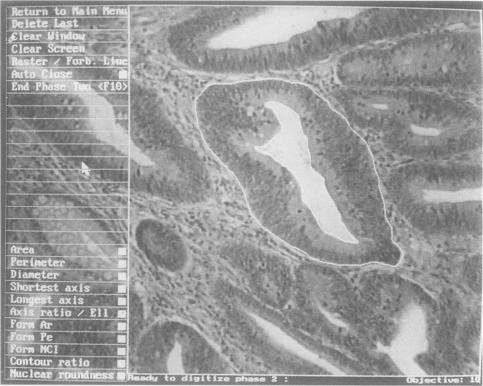
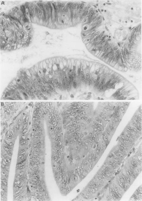
Mitotic counts were conducted in tissue sections of 59 colorectal adenomas. Of these, 20 showed mild, 20 moderate, and 19 severe dysplasia, according to blind duplicate assessments by two pathologists. Forty three were classified as tubular adenomas and 16 as "villous" adenomas (tubulo-villous and villous). The number of mitoses, both per unit area of epithelium (area weighted mitotic counts, AWMC) and per colonic crypt (mitotic counts per colonic crypt MCCC), was scored in the most dysplastic area within the adenoma. Mitotic figures were counted using a light microscope (ocular x 10, objective x 40, NA 0.75), and the area of the glandular epithelium was measured using an interactive video overlay measurement system. Twenty glands per specimen were assessed. In the intra-observer reproducibility tests, the coefficients of error for the AWMC and MCCC were 4.5% and 7.4% respectively.
For the AWMC a significant difference was found between mild and moderate as well as between mild and severe dysplasia, but not between moderate and severe dysplasia. The results of the MCCC showed the same trend, but the differences did not reach a significant level. Furthermore, cases classified as mild dysplasia were found that showed numerous mitoses, while cases classified as severe dysplasia were found with only very few mitoses. No significant difference in AWMC was found between tubular and villous adenomas. Thus the different malignant potential of tubular and villous adenomas was not reflected by a difference in AWMC. A seemingly strong difference for MCCC between tubular and "villous" adenomas appeared to depend completely on the difference in crypt size between these two groups.
The area weighted mitotic count, rather than the mitotic count per colonic crypt, may be useful for assessing the proliferation rate in colorectal adenomas.
肿瘤的增殖活性反映其恶性潜能。在结直肠腺瘤中,对有丝分裂数量的主观印象是常用于评估发育异常程度的一个标准。由于这些关于有丝分裂活性的主观印象可能缺乏可重复性,本研究的目的是进行客观分析。
对59例结直肠腺瘤的组织切片进行有丝分裂计数。其中,根据两位病理学家的双盲重复评估,20例显示轻度发育异常,20例为中度,19例为重度。43例被分类为管状腺瘤,16例为“绒毛状”腺瘤(管状绒毛状和绒毛状)。在腺瘤内发育异常最严重的区域,对每单位上皮面积的有丝分裂数量(面积加权有丝分裂计数,AWMC)和每个结肠隐窝的有丝分裂数量(每个结肠隐窝有丝分裂计数,MCCC)进行评分。使用光学显微镜(目镜×10,物镜×40,数值孔径0.75)计数有丝分裂象,并使用交互式视频叠加测量系统测量腺上皮的面积。每个标本评估20个腺体。在观察者内重复性测试中,AWMC和MCCC的误差系数分别为4.5%和7.4%。
对于AWMC,在轻度与中度以及轻度与重度发育异常之间发现有显著差异,但在中度与重度发育异常之间未发现显著差异。MCCC的结果显示出相同趋势,但差异未达到显著水平。此外,发现分类为轻度发育异常的病例有大量有丝分裂,而分类为重度发育异常的病例只有很少的有丝分裂。在管状腺瘤和绒毛状腺瘤之间,AWMC未发现显著差异。因此,管状腺瘤和绒毛状腺瘤不同的恶性潜能未通过AWMC的差异反映出来。管状腺瘤和“绒毛状”腺瘤之间MCCC看似强烈的差异似乎完全取决于这两组之间隐窝大小的差异。
面积加权有丝分裂计数,而非每个结肠隐窝的有丝分裂计数,可能有助于评估结直肠腺瘤的增殖率。