Michel T, Li G K, Busconi L
Cardiovascular Division, Brigham and Women's Hospital, Harvard Medical School, Boston, MA 02115.
Proc Natl Acad Sci U S A. 1993 Jul 1;90(13):6252-6. doi: 10.1073/pnas.90.13.6252.
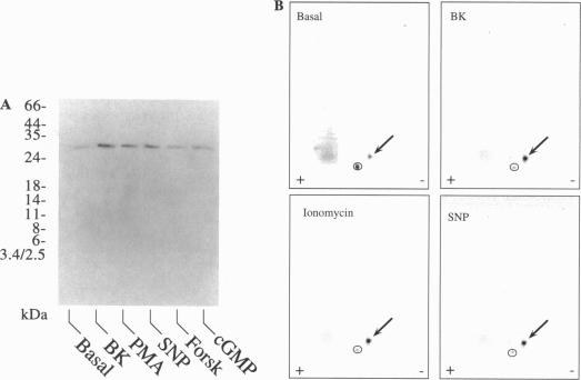
In the vascular endothelium, diverse cell surface receptors are coupled to the Ca2+/calmodulin-dependent activation of nitric oxide (NO) synthase. We now report that, in intact cultured endothelial cells, several drugs and agonists are associated with increased serine phosphorylation of the endothelial NO synthase. We biosynthetically labeled bovine aortic endothelial cells with [32P]orthophosphoric acid, exposed the cells to various drugs and hormones, and then immunoprecipitated the enzyme from cell extracts using a highly specific anti-peptide antibody. The marked endothelial NO synthase phosphorylation induced by bradykinin is maximal only after 5 min of agonist exposure and is stable for at least 20 min. Basal and agonist-induced phosphorylation of the NO synthase in endothelial cells is completely inhibited by the calmodulin antagonist compound W-7. We prepared subcellular fractions of endothelial cells that had been biosynthetically labeled with [35S]methionine or [32P]orthophosphoric acid and immunoprecipitated the endothelial NO synthase from untreated (basal) and bradykinin-treated cells. In the basal state, [35S]methionine-labeled endothelial NO synthase is associated primarily with the particulate cellular fraction, but the phosphorylated enzyme is primarily cytosolic. Following exposure to bradykinin, a substantial fraction of the [35S]methionine-labeled NO synthase is now found in the cytosolic fraction, associated with a marked increase in the level of cytosolic enzyme phosphorylation. We propose that agonist-induced phosphorylation of NO synthase is associated with translocation of the enzyme from membrane to cytosol and may thereby regulate the biological effects of endothelial NO synthesis in situ.
在血管内皮细胞中,多种细胞表面受体与一氧化氮(NO)合酶的Ca2+/钙调蛋白依赖性激活相偶联。我们现在报告,在完整的培养内皮细胞中,几种药物和激动剂与内皮型NO合酶丝氨酸磷酸化增加有关。我们用[32P]正磷酸对牛主动脉内皮细胞进行生物合成标记,将细胞暴露于各种药物和激素中,然后使用高度特异性的抗肽抗体从细胞提取物中免疫沉淀该酶。缓激肽诱导的显著内皮型NO合酶磷酸化仅在激动剂暴露5分钟后达到最大值,并至少稳定20分钟。钙调蛋白拮抗剂化合物W-7完全抑制内皮细胞中NO合酶的基础磷酸化和激动剂诱导的磷酸化。我们制备了用[35S]甲硫氨酸或[32P]正磷酸进行生物合成标记的内皮细胞亚细胞组分,并从未经处理(基础)和缓激肽处理的细胞中免疫沉淀内皮型NO合酶。在基础状态下,[35S]甲硫氨酸标记的内皮型NO合酶主要与细胞颗粒部分相关,但磷酸化的酶主要位于胞质中。暴露于缓激肽后,现在在胞质组分中发现了相当一部分[35S]甲硫氨酸标记的NO合酶,这与胞质酶磷酸化水平的显著增加相关。我们提出,激动剂诱导的NO合酶磷酸化与该酶从膜向胞质的转位有关,从而可能原位调节内皮NO合成的生物学效应。