Swerdlow S H, Saboorian M H, Pelstring R J, Williams M E
Department of Pathology, University of Cincinnati College of Medicine, Ohio.
Am J Pathol. 1993 Jan;142(1):329-37.
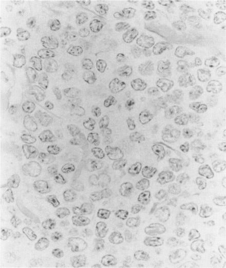
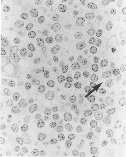
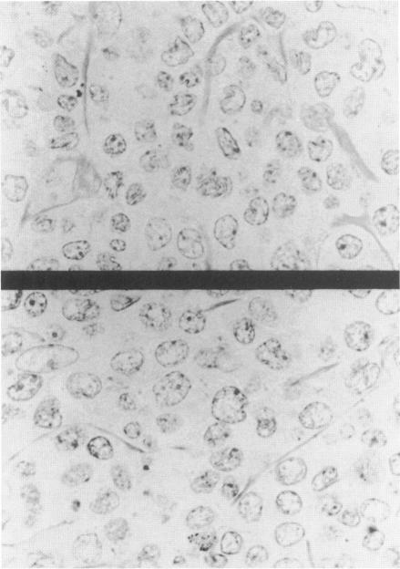
Centrocytic lymphoma (ML,CC) is distinguished from other cleaved follicular center cell (FCC) lymphomas in the Kiel classification by the lack of noncleaved FCC and not by morphological differences between the centrocytes (cleaved cells). Immunophenotypic and genotypic studies, however, have shown that the centrocytes in ML,CC are distinct from those of other small cleaved FCC lymphomas (ML,FCC,SC). To morphologically compare the cells of ML,CC with nine previously studied ML,FCC,SC and to relate the findings in ML,CC to the varied descriptions of lymphomas of intermediate differentiation, a morphometric analysis of 22 ML,CC was performed. Nuclei in ML,CC were, on average, significantly larger, rounder, and had less frequent nucleoli than those in ML,FCC,SC; however, the proportion of small round lymphocytes did not differ. Among the ML,CC, the only apparent immunophenotypic/genotypic correlate that was identified was greater nuclear ellipticity for the biopsies lacking chromosome 11q13 bcl-1 or PRAD1 rearrangement. Repeat biopsies in four patients with ML,CC showed an increase in nuclear size. These data demonstrate that a lack of transformed cells is not the only morphological difference between ML,CC and ML,FCC,SC. The morphological distinction, however, is not based on the proportion of small round lymphocytes present. In addition, the morphometric parameters illustrate the nuclear variability among ML,CC and demonstrate how the disease may evolve over time.
中心细胞性淋巴瘤(ML,CC)在基尔分类中与其他裂核滤泡中心细胞(FCC)淋巴瘤的区别在于缺乏未裂核FCC,而非中心细胞(裂核细胞)之间的形态学差异。然而,免疫表型和基因分型研究表明,ML,CC中的中心细胞与其他小裂核FCC淋巴瘤(ML,FCC,SC)中的中心细胞不同。为了在形态学上比较ML,CC的细胞与之前研究的9种ML,FCC,SC,并将ML,CC的研究结果与中间分化淋巴瘤的各种描述相关联,对22例ML,CC进行了形态计量分析。ML,CC中的细胞核平均而言明显更大、更圆,核仁频率更低,比ML,FCC,SC中的细胞核;然而,小圆形淋巴细胞的比例没有差异。在ML,CC中,唯一确定的明显免疫表型/基因分型相关性是缺乏11q13染色体bcl-1或PRAD1重排的活检标本中核椭圆率更高。4例ML,CC患者的重复活检显示核大小增加。这些数据表明,缺乏转化细胞并不是ML,CC和ML,FCC,SC之间唯一的形态学差异。然而,形态学差异并非基于小圆形淋巴细胞的比例。此外,形态计量参数说明了ML,CC中核的变异性,并证明了该疾病可能如何随时间演变。