• 文献检索
  • 文档翻译
  • 深度研究
  • 学术资讯
  • Suppr Zotero 插件Zotero 插件
  • 邀请有礼
  • 套餐&价格
  • 历史记录
应用&插件
Suppr Zotero 插件Zotero 插件浏览器插件Mac 客户端Windows 客户端微信小程序
定价
高级版会员购买积分包购买API积分包
服务
文献检索文档翻译深度研究API 文档MCP 服务
关于我们
关于 Suppr公司介绍联系我们用户协议隐私条款
关注我们

Suppr 超能文献

核心技术专利:CN118964589B侵权必究
粤ICP备2023148730 号-1Suppr @ 2026

文献检索

告别复杂PubMed语法,用中文像聊天一样搜索,搜遍4000万医学文献。AI智能推荐,让科研检索更轻松。

立即免费搜索

文件翻译

保留排版,准确专业,支持PDF/Word/PPT等文件格式,支持 12+语言互译。

免费翻译文档

深度研究

AI帮你快速写综述,25分钟生成高质量综述,智能提取关键信息,辅助科研写作。

立即免费体验

视网膜和视神经乳头灌注的二维映射。

Two dimensional mapping of the perfusion of the retina and optic nerve head.

作者信息

Michelson G, Schmauss B

机构信息

Department of Ophthalmology, University Erlangen-Nürnberg, Germany.

出版信息

Br J Ophthalmol. 1995 Dec;79(12):1126-32. doi: 10.1136/bjo.79.12.1126.

DOI:10.1136/bjo.79.12.1126
PMID:8562550
原文链接:https://pmc.ncbi.nlm.nih.gov/articles/PMC505355/
Abstract

AIM

To present a new non-invasive method of performing a high definition topography of perfused vessels of the retina and the optic nerve head with simultaneous evaluation of blood flow.

METHOD

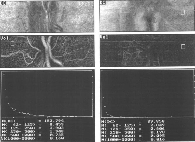
By a combination of a laser Doppler flowmeter with a scanning laser system the perfusion of the retina and the optic nerve head is visualised. The principles of measuring blood flow by laser Doppler flowmetry are based on the optical Doppler effect: laser light scattered by a moving particle is shifted in frequency by an amount delta f. Our data acquisition and evaluation system is a modified laser scanning tomograph. The technical data are retinal area of measurement 2.7 mm x 0.7 mm, 10 degrees field with 256 points x 64 lines, measurement accuracy 10 microns, wavelength 670 nm and 790 nm, light power 100 microW and 200 microW, data acquisition time 2.048 s. Every line is scanned 128 times by a line sampling rate of 4000 Hz. By performing a discrete fast Fourier transformation over 128 intensities of each retinal point the laser Doppler shift is calculated for each retinal point. With these data a two dimensional map with 256 x 64 points of the retinal perfusion is created. The brightness of the pixel is coded by the value of the Doppler shift. Offline capillary blood flow is estimated in arbitrary units according to the theory of laser Doppler flowmetry in every region of interest of the perfusion picture. We estimated the reliability and the validity of the method. Retinal blood flow was measured by scanning laser Doppler flowmetry (SLDF) while varying intraocular pressure by a suction cup of three healthy volunteers. Measurements of retinal blood flow performed in 47 eyes by the presented method (SLDF) were correlated with data gained by a commercially available laser Doppler flowmeter. Perfusion pictures of the superficial retinal layer and of deep prelaminar layers in the optic nerve head are presented.

RESULTS

The reliability coefficients r1 of 'flow', 'volume', and 'velocity' were 0.84, 0.85, and 0.84 respectively. We found a significant linear relation between SLDF flow and the ocular perfusion pressure (r = 0.84, p < 0.001). Comparative measurements of the retinal blood flow by SLDF and a commercially available laser Doppler flowmeter showed a linear and significant relation (flow r = 0.6, p < 0.0001, volume r = 0.4, p < 0.01). Capillaries of the retinal superficial vasculature or deep ciliary sourced capillaries of the optic nerve head became visible with a high resolution by the confocal technique dependent on the focus. Offline, the blood flow variables of areas of 100 microns x 100 microns were calculated.

CONCLUSION

SLDF enables the visualisation of perfused capillaries and vessels of the retina and the optic nerve head in high resolution by two dimensional mapping of perfusion variables which are encoded by the Doppler signal. This method achieves simultaneously qualitative and quantitative evaluation of capillary blood flow of distinct areas of the capillary meshwork.

https://cdn.ncbi.nlm.nih.gov/pmc/blobs/3f30/505355/bd1956d45f57/brjopthal00024-0068-a.jpg
https://cdn.ncbi.nlm.nih.gov/pmc/blobs/3f30/505355/3d454d0b4eb8/brjopthal00024-0067-a.jpg
https://cdn.ncbi.nlm.nih.gov/pmc/blobs/3f30/505355/27ea7ab1269c/brjopthal00024-0067-b.jpg
https://cdn.ncbi.nlm.nih.gov/pmc/blobs/3f30/505355/bd1956d45f57/brjopthal00024-0068-a.jpg
https://cdn.ncbi.nlm.nih.gov/pmc/blobs/3f30/505355/3d454d0b4eb8/brjopthal00024-0067-a.jpg
https://cdn.ncbi.nlm.nih.gov/pmc/blobs/3f30/505355/27ea7ab1269c/brjopthal00024-0067-b.jpg
https://cdn.ncbi.nlm.nih.gov/pmc/blobs/3f30/505355/bd1956d45f57/brjopthal00024-0068-a.jpg
摘要

目的

提出一种新的非侵入性方法,用于对视网膜和视神经乳头的灌注血管进行高分辨率地形图绘制,并同时评估血流情况。

方法

通过将激光多普勒血流仪与扫描激光系统相结合,可实现视网膜和视神经乳头灌注情况的可视化。激光多普勒血流测量法测量血流的原理基于光学多普勒效应:运动粒子散射的激光在频率上会发生Δf量的偏移。我们的数据采集和评估系统是一台经过改进的激光扫描断层扫描仪。技术参数为:视网膜测量面积2.7毫米×0.7毫米,10度视野,256点×64线,测量精度10微米,波长670纳米和790纳米,光功率100微瓦和200微瓦,数据采集时间2.048秒。每条线以4000赫兹的线采样率扫描128次。通过对每个视网膜点的128个强度值进行离散快速傅里叶变换,计算出每个视网膜点的激光多普勒频移。利用这些数据创建一个256×64点的视网膜灌注二维图。像素的亮度由多普勒频移值编码。根据激光多普勒血流测量理论,以任意单位离线估计灌注图像每个感兴趣区域的毛细血管血流。我们评估了该方法的可靠性和有效性。在三名健康志愿者中,通过吸盘改变眼内压的同时,用扫描激光多普勒血流测量法(SLDF)测量视网膜血流。用所提出的方法(SLDF)对47只眼睛进行的视网膜血流测量结果与商用激光多普勒血流仪获得的数据进行了相关性分析。展示了视网膜表层和视神经乳头深层板层前的灌注图像。

结果

“流量”“体积”和“速度”的可靠性系数r1分别为0.84、0.85和0.84。我们发现SLDF流量与眼灌注压之间存在显著的线性关系(r = 0.84,p < 0.001)。SLDF与商用激光多普勒血流仪对视网膜血流的比较测量显示出线性且显著的关系(流量r = 0.6,p < 0.0001,体积r = 0.4,p < 0.01)。通过共聚焦技术,根据焦点不同,视网膜浅层脉管系统的毛细血管或视神经乳头深层睫状来源的毛细血管能够以高分辨率清晰可见。离线计算100微米×100微米区域的血流变量。

结论

SLDF通过对由多普勒信号编码的灌注变量进行二维映射,能够高分辨率地可视化视网膜和视神经乳头的灌注毛细血管和血管。该方法可同时对毛细血管网不同区域的毛细血管血流进行定性和定量评估。

相似文献

1
Two dimensional mapping of the perfusion of the retina and optic nerve head.视网膜和视神经乳头灌注的二维映射。
Br J Ophthalmol. 1995 Dec;79(12):1126-32. doi: 10.1136/bjo.79.12.1126.
2
[2-dimensional mapping and retinal and papillary microcirculation using scanning laser Doppler flowmetry].[使用扫描激光多普勒血流仪进行二维映射以及视网膜和乳头体微循环研究]
Klin Monbl Augenheilkd. 1995 Sep;207(3):180-90. doi: 10.1055/s-2008-1035365.
3
Clinical investigation of the combination of a scanning laser ophthalmoscope and laser Doppler flowmeter.扫描激光检眼镜与激光多普勒血流仪联合应用的临床研究
Ger J Ophthalmol. 1995 Nov;4(6):342-9.
4
Principle, validity, and reliability of scanning laser Doppler flowmetry.扫描激光多普勒血流仪的原理、有效性和可靠性。
J Glaucoma. 1996 Apr;5(2):99-105.
5
Automatic full field analysis of perfusion images gained by scanning laser Doppler flowmetry.通过扫描激光多普勒血流仪获得的灌注图像的自动全场分析。
Br J Ophthalmol. 1998 Nov;82(11):1294-300. doi: 10.1136/bjo.82.11.1294.
6
Perfusion of the juxtapapillary retina and optic nerve head in acute ocular hypertension.急性高眼压状态下视乳头旁视网膜及视神经乳头的灌注情况
Ger J Ophthalmol. 1996 Nov;5(6):315-21.
7
Effect of breathing 100% oxygen on retinal and optic nerve head capillary blood flow in smokers and non-smokers.吸入100%氧气对吸烟者和非吸烟者视网膜及视神经乳头毛细血管血流的影响。
Br J Ophthalmol. 1997 May;81(5):365-9. doi: 10.1136/bjo.81.5.365.
8
Reproducibility of retinal and optic nerve head blood flow measurements with scanning laser Doppler flowmetry.扫描激光多普勒血流仪测量视网膜和视神经乳头血流的可重复性。
J Glaucoma. 1997 Jun;6(3):157-64.
9
Functional imaging of the retinal microvasculature by scanning laser Doppler flowmetry.通过扫描激光多普勒血流仪对视网膜微血管进行功能成像。
Int Ophthalmol. 2001;23(4-6):327-35. doi: 10.1023/a:1014402730503.
10
Perfusion of the juxtapapillary retina and the neuroretinal rim area in primary open angle glaucoma.原发性开角型青光眼患者视乳头旁视网膜及神经视网膜边缘区域的灌注情况
J Glaucoma. 1996 Apr;5(2):91-8.

引用本文的文献

1
Arterial Hypertension and the Hidden Disease of the Eye: Diagnostic Tools and Therapeutic Strategies.动脉高血压与眼睛的隐匿性疾病:诊断工具与治疗策略。
Nutrients. 2022 May 25;14(11):2200. doi: 10.3390/nu14112200.
2
Imaging video plethysmography shows reduced signal amplitude in glaucoma patients in the area of the microvascular tissue of the optic nerve head.影像视频体积描记法显示,青光眼患者视神经头微血管组织区域的信号幅度降低。
Graefes Arch Clin Exp Ophthalmol. 2021 Feb;259(2):483-494. doi: 10.1007/s00417-020-04934-y. Epub 2020 Sep 22.
3
Adaptive optics imaging of the human retina.

本文引用的文献

1
Simultaneous measurement of ocular micro- and macrocirculation, intraocular pressure, and systemic functions.同时测量眼部微循环和大循环、眼压及全身功能。
Ger J Ophthalmol. 1994 Jan;3(1):48-53.
2
Reactions following intravenous fluorescein.静脉注射荧光素后的反应。
Am J Ophthalmol. 1971 Nov;72(5):861-8. doi: 10.1016/0002-9394(71)91681-3.
3
Retinal capillary blood flow measurement with a scanning laser ophthalmoscope. Preliminary results.使用扫描激光检眼镜测量视网膜毛细血管血流。初步结果。
自适应光学视网膜成像。
Prog Retin Eye Res. 2019 Jan;68:1-30. doi: 10.1016/j.preteyeres.2018.08.002. Epub 2018 Aug 27.
4
Diurnal variations and pulpal status: is there a need for FFT besides LDF?昼夜节律变化和牙髓状态:除了 LDF 之外,FFT 是否有必要?
Lasers Med Sci. 2018 Dec;33(9):1891-1900. doi: 10.1007/s10103-018-2550-5. Epub 2018 Jun 13.
5
Retinal blood flow is increased in type 1 diabetes mellitus patients with advanced stages of retinopathy.在患有晚期视网膜病变的1型糖尿病患者中,视网膜血流量会增加。
BMC Endocr Disord. 2016 May 26;16(1):25. doi: 10.1186/s12902-016-0105-y.
6
Visualization of retinal vascular structure and perfusion with a nonconfocal adaptive optics scanning light ophthalmoscope.使用非共焦自适应光学扫描激光检眼镜对视网膜血管结构和灌注进行可视化。
J Opt Soc Am A Opt Image Sci Vis. 2014 Mar 1;31(3):569-79. doi: 10.1364/JOSAA.31.000569.
7
Pilot study for the evaluation of morphological and functional changes in retinal blood flow in patients with insulin resistance and/or type 2 diabetes mellitus.评估胰岛素抵抗和/或2型糖尿病患者视网膜血流形态和功能变化的初步研究。
J Diabetes Sci Technol. 2012 Jan 1;6(1):163-8. doi: 10.1177/193229681200600120.
8
Altered temporal peripapillary retinal flow in patients with disc hemorrhages.盘出血患者的视盘周围视网膜 temporal 血流改变。
Graefes Arch Clin Exp Ophthalmol. 2010 Dec;248(12):1771-5. doi: 10.1007/s00417-010-1441-7. Epub 2010 Jul 16.
9
Inability of a Confocal Scanning Laser Doppler Flowmeter to Measure Choroidal Blood Flow in the Pig Eye.共焦扫描激光多普勒血流仪无法测量猪眼脉络膜血流
Open Ophthalmol J. 2008 Nov 14;2:146-52. doi: 10.2174/1874364100802010146.
10
A placebo-controlled study of retinal blood flow changes by pentoxifylline and metabolites in humans.一项关于己酮可可碱及其代谢产物对人体视网膜血流变化影响的安慰剂对照研究。
Br J Clin Pharmacol. 2006 Feb;61(2):138-47. doi: 10.1111/j.1365-2125.2005.02549.x.
Ophthalmology. 1991 Jun;98(6):996-1000. doi: 10.1016/s0161-6420(91)32192-4.
4
Laser Doppler flowmetry in the optic nerve.视神经中的激光多普勒血流仪
Exp Eye Res. 1992 Sep;55(3):499-506. doi: 10.1016/0014-4835(92)90123-a.