Svanberg K, Liu D L, Wang I, Andersson-Engels S, Stenram U, Svanberg S
Department of Oncology, Lund University Hospital, Sweden.
Br J Cancer. 1996 Nov;74(10):1526-33. doi: 10.1038/bjc.1996.584.
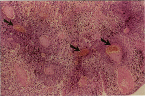
The efficacy of photodynamic therapy (PDT) using delta-aminolaevulinic acid (ALA)-induced protoporphyrin IX (PpIX) sensitisation and laser light at 635 nm was investigated in the treatment of experimental hepatic tumours. The model of liver tumours was induced either by local inoculation or by administration of tumour cells through the portal vein in rats. ALA at a dose of 60 mg kg(-1) b.w. was intravenously administered 60 min before PDT. PpIX accumulation in tumour, normal liver and abdominal wall muscle was detected by means of laser-induced fluorescence (LIF). Laser Doppler imaging (LDI) was used to determine changes in the superficial blood flow in connection with PDT. Histopathological examinations were performed to evaluate the PDT effects on the tumour and the surrounding liver tissue, including pathological features in the microvascular system. The accumulation of PpIX, as monitored by LIF, showed high fluorescence intensities at about 635 nm in both the hepatic tumour tissue and normal liver and low values in the abdominal wall. LDI demonstrated that the blood flow in the treated tumour and its surrounding normal liver tissue decreased immediately after the PDT, indicating an effect on the vascular system. A large number of thrombi in the irradiated tumour were found microscopically 3 h after the PDT. The tumour growth rate showed a marked decrease when evaluated 3 and 6 days after the treatment. These results show that the ALA-PDT is effective in the inhibition of growth of experimental hepatic tumours.
研究了使用δ-氨基乙酰丙酸(ALA)诱导原卟啉IX(PpIX)敏化并结合635 nm激光进行光动力疗法(PDT)治疗实验性肝肿瘤的疗效。肝肿瘤模型通过在大鼠局部接种或经门静脉注射肿瘤细胞诱导建立。在PDT前60分钟静脉注射剂量为60 mg kg(-1)体重的ALA。通过激光诱导荧光(LIF)检测肿瘤、正常肝脏和腹壁肌肉中PpIX的积累。使用激光多普勒成像(LDI)确定与PDT相关的浅表血流变化。进行组织病理学检查以评估PDT对肿瘤和周围肝组织的影响,包括微血管系统的病理特征。通过LIF监测,PpIX的积累在肝肿瘤组织和正常肝脏中均在约635 nm处显示出高荧光强度,而在腹壁中荧光值较低。LDI表明,PDT后处理的肿瘤及其周围正常肝组织中的血流立即减少,表明对血管系统有影响。PDT后3小时在显微镜下发现照射肿瘤中有大量血栓。在治疗后3天和6天评估时,肿瘤生长速率显著降低。这些结果表明,ALA-PDT在抑制实验性肝肿瘤生长方面是有效的。