Culora G A, Ramsay A D, Theaker J M
Department of Histopathology, Southampton University Hospitals.
J Clin Pathol. 1996 Oct;49(10):844-7. doi: 10.1136/jcp.49.10.844.
To alert pathologists to the spectrum of histological appearances that may be seen in injection site reactions related to aluminium.
Four cases of injection site reaction were examined microscopically using routine staining with haematoxylin and eosin, electron microscopy and by electron probe microanalysis.
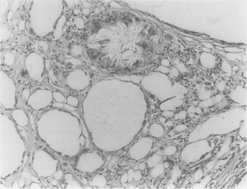
As in previous reports, all four cases included collections of histiocytes which contained faint granular brownish refractile material within their cytoplasm; ultrastructural examination showed this to be aluminium. Two cases showed a prominent inflammatory reaction with numerous lymphoid follicles and a notable eosinophilic infiltrate. Two cases showed unusual features not described previously. In one, there was a sclerosing lipogranuloma-like reaction with unlined cystic spaces containing crystalline material. The other case presented as a large symptomatic subcutaneous swelling which microscopically showed diffuse and wide-spread involvement of the subcutis by a lymphoid infiltrate with prominent lymphoid follicles.
This report highlights the changes encountered in aluminium injection site reactions and emphasises that the lesions have a wider range of histological appearances than described previously.
提醒病理学家注意与铝相关的注射部位反应可能出现的一系列组织学表现。
对4例注射部位反应进行显微镜检查,采用苏木精和伊红常规染色、电子显微镜检查以及电子探针微分析。
与之前的报道一样,所有4例均有组织细胞聚集,其细胞质内含有微弱的颗粒状褐色折光物质;超微结构检查显示这是铝。2例表现为显著的炎症反应,有大量淋巴滤泡和明显的嗜酸性粒细胞浸润。2例表现出先前未描述的异常特征。其中1例为硬化性脂肉芽肿样反应,有无内衬的囊腔,内含结晶物质。另一例表现为有症状的巨大皮下肿胀,显微镜下显示皮下组织弥漫性广泛受累,有淋巴细胞浸润和明显的淋巴滤泡。
本报告突出了铝注射部位反应中遇到的变化,并强调这些病变的组织学表现范围比先前描述的更广。