Gentile R C, Liebmann J M, Tello C, Stegman Z, Weissman S S, Ritch R
Ocular Imaging Center, New York Eye and Ear Infirmary, New York 10003, USA.
Br J Ophthalmol. 1996 Oct;80(10):895-9. doi: 10.1136/bjo.80.10.895.
Acute anterior uveitis has diverse causes and systemic associations. Inflammation is predominantly localised to the iris and pars plicata. Little is known about the in vivo effects of uveitis on ciliary body anatomy.
Bilateral, high frequency, high resolution, ultrasound biomicroscopy was performed on consecutive patients with unilateral anterior uveitis to evaluate ciliary body anatomy. Imaging was repeated when possible during the clinical course. The cross sectional area of the anterior ciliary body was measured using image processing and analysis software. Measurements from the uveitic eyes were compared with the fellow eyes and the effect of treatment was evaluated.
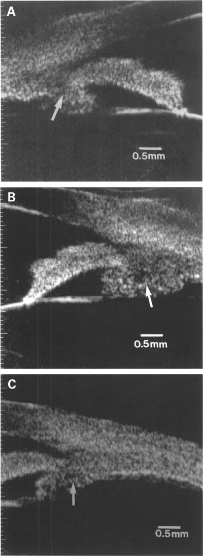
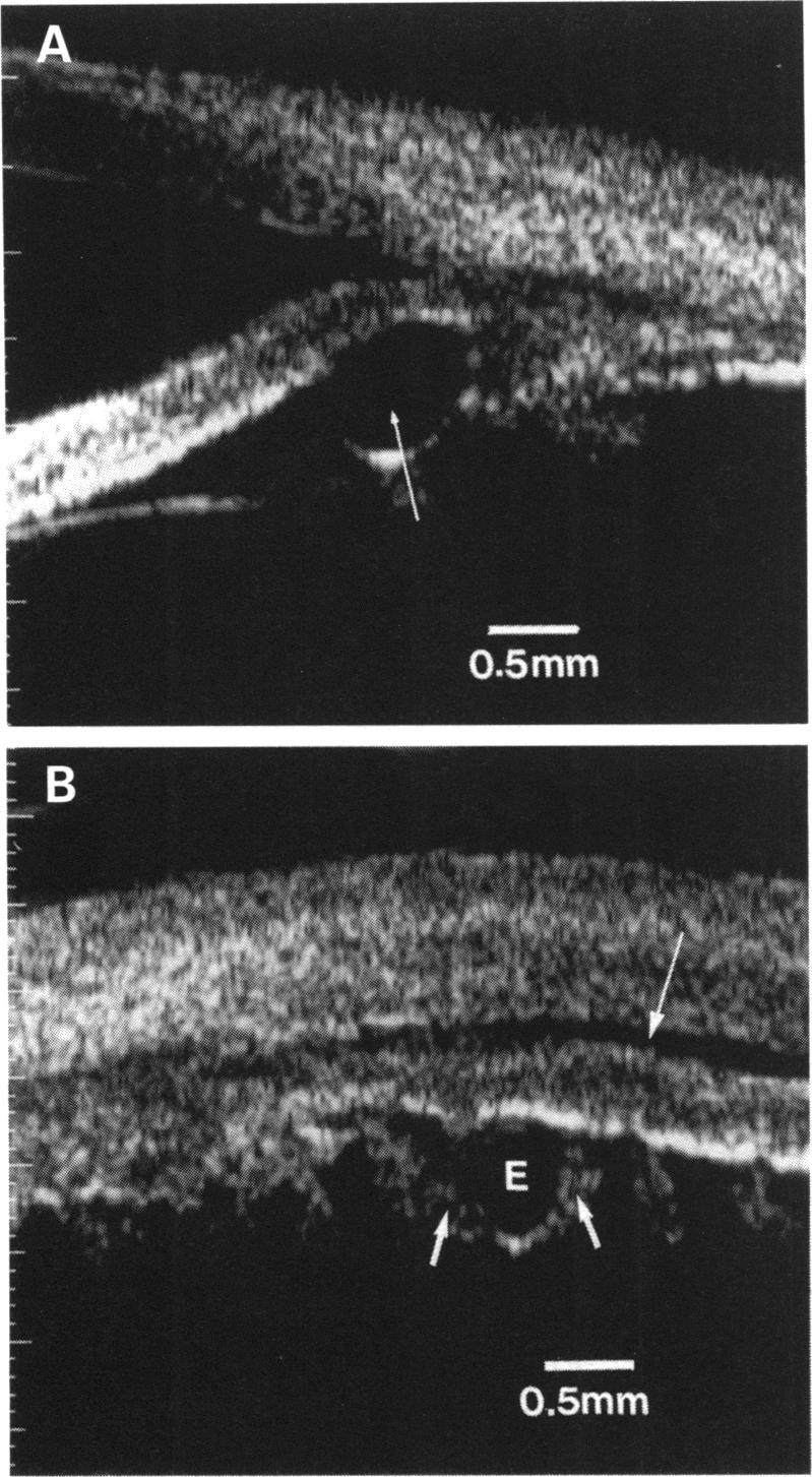
Fourteen patients were enrolled. Ultrasound biomicroscopy demonstrated a larger ciliary body cross sectional area in the uveitic eyes compared with the fellow, clinically uninvolved eyes (2.45 (SD 0.48) mm2 versus 1.55 (SD 0.15) mm2, (p = 0.0000; paired t test)). A ciliochoroidal effusion was present in one uveitic eye. Epithelial cysts were imaged bilaterally in four uveitic patients (29%) and unilaterally in unaffected eyes of two uveitic patients. Ciliary body cross sectional area decreased following steroid therapy (p = 0.0001; paired t test). New cysts were noted in three uveitic eyes during the follow up period and in none of the fellow, unaffected eyes.
Ultrasound biomicroscopy offers a new approach to the evaluation of anterior uveitis. The response to treatment can be evaluated objectively and therapeutic efficacy can be more easily assessed. It has the potential to help elucidate the pathophysiology and anatomical changes of this heterogeneous group of disorders.
急性前葡萄膜炎病因多样且与全身情况相关。炎症主要局限于虹膜和睫状体扁平部。关于葡萄膜炎对睫状体解剖结构的体内影响知之甚少。
对连续性单侧前葡萄膜炎患者进行双侧、高频、高分辨率超声生物显微镜检查以评估睫状体解剖结构。在临床过程中尽可能重复成像。使用图像处理和分析软件测量前睫状体的横截面积。将患眼的测量结果与对侧眼进行比较,并评估治疗效果。
纳入14例患者。超声生物显微镜检查显示,与对侧临床未受累眼相比,患眼的睫状体横截面积更大(2.45(标准差0.48)mm²对1.55(标准差0.15)mm²,(p = 0.0000;配对t检验))。一只患眼中存在睫脉络膜积液。4例(29%)葡萄膜炎患者双侧可见上皮囊肿,2例葡萄膜炎患者未受累眼单侧可见上皮囊肿。类固醇治疗后睫状体横截面积减小(p = 0.0001;配对t检验)。随访期间3只患眼中发现新囊肿,对侧未受累眼中均未发现。
超声生物显微镜检查为前葡萄膜炎的评估提供了一种新方法。可客观评估对治疗的反应,更易于评估治疗效果。它有可能有助于阐明这类异质性疾病的病理生理学和解剖学变化。