Zhou D, Birkenmeier C S, Williams M W, Sharp J J, Barker J E, Bloch R J
Department of Physiology, University of Maryland School of Medicine, Baltimore 21201, USA.
J Cell Biol. 1997 Feb 10;136(3):621-31. doi: 10.1083/jcb.136.3.621.
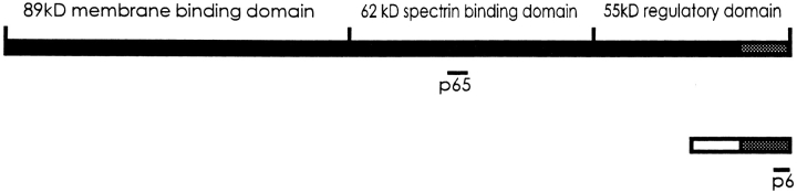
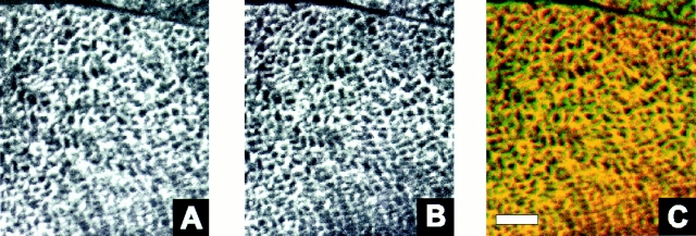
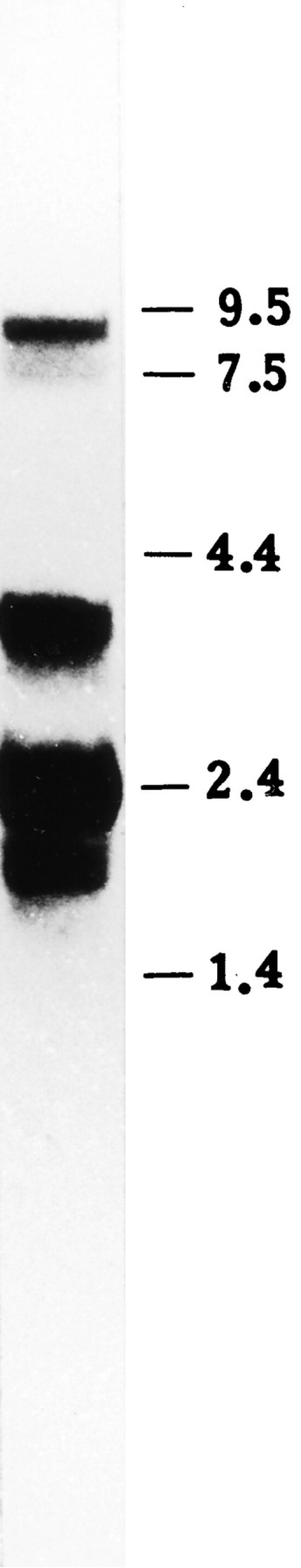
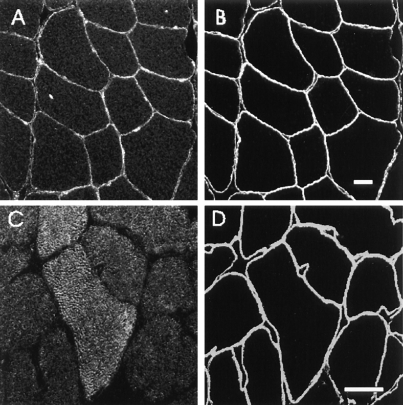
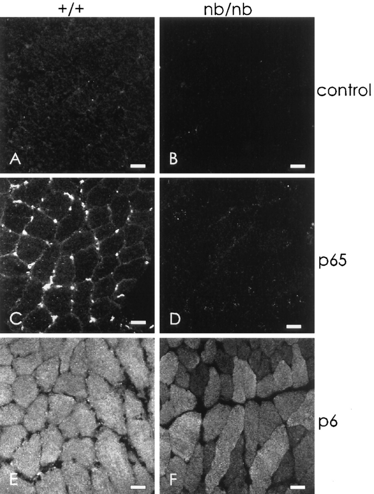
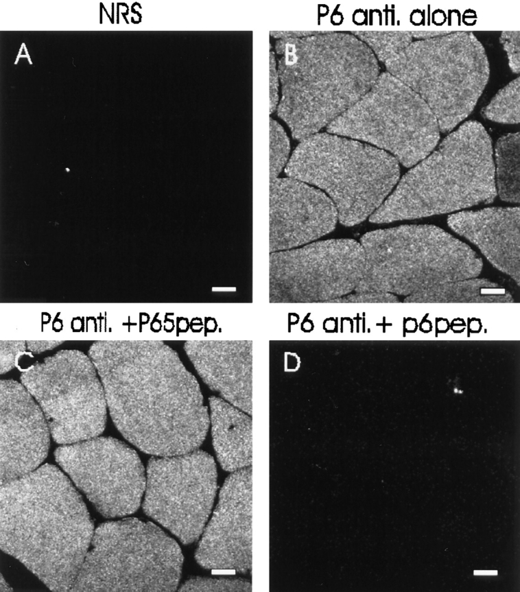
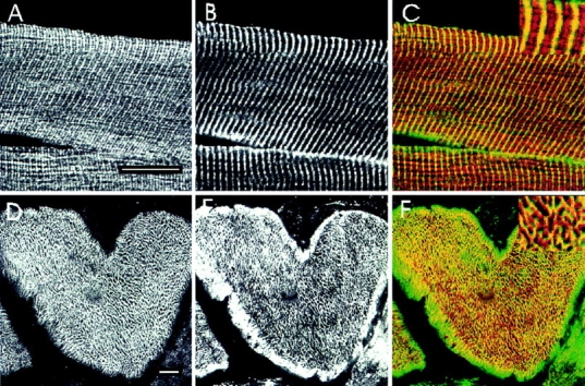
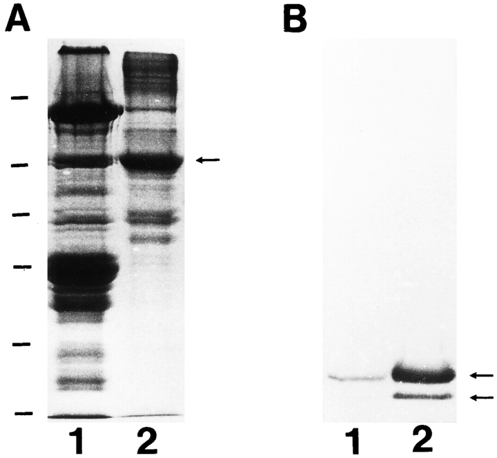
We have recently found that the erythroid ankyrin gene, Ank1, expresses isoforms in mouse skeletal muscle, several of which share COOH-terminal sequence with previously known Ank1 isoforms but have a novel, highly hydrophobic 72-amino acid segment at their NH2 termini. Here, through the use of domain-specific peptide antibodies, we report the presence of the small ankyrins in rat and rabbit skeletal muscle and demonstrate their selective association with the sarcoplasmic reticulum. In frozen sections of rat skeletal muscle, antibodies to the spectrin-binding domain (anti-p65) react only with a 210-kD Ank1 and label the sarcolemma and nuclei, while antibodies to the COOH terminus of the small ankyrin (anti-p6) react with peptides of 20 to 26 kD on immunoblots and decorate the myoplasm in a reticular pattern. Mice homozygous for the normoblastosis mutation (gene symbol nb) are deficient in the 210-kD ankyrin but contain normal levels of the small ankyrins in the myoplasm. In nb/nb skeletal muscle, anti-p65 label is absent from the sarcolemma, whereas anti-p6 label shows the same distribution as in control skeletal muscle. In normal skeletal muscle of the rat, anti-p6 decorates Z lines, as defined by antidesmin distribution, and is also present at M lines where it surrounds the thick myosin filaments. Immunoblots of the proteins isolated with rabbit sarcoplasmic reticulum indicate that the small ankyrins are highly enriched in this fraction. When expressed in transfected HEK 293 cells, the small ankyrins are distributed in a reticular pattern resembling the ER if the NH2-terminal hydrophobic domain is present, but they are uniformly distributed in the cytosol if this domain is absent. These results suggest that the small ankyrins are integral membrane proteins of the sarcoplasmic reticulum. We propose that, unlike the 210-kD form of Ank1, previously localized to the sarcolemma and believed to be a part of the supporting cytoskeleton, the small Ank1 isoforms may stabilize the sarcoplasmic reticulum by linking it to the contractile apparatus.
我们最近发现,红细胞锚蛋白基因Ank1在小鼠骨骼肌中表达多种同工型,其中几种在COOH末端序列上与先前已知的Ank1同工型相同,但在其NH2末端有一个新的、高度疏水的72个氨基酸的片段。在这里,通过使用结构域特异性肽抗体,我们报告了大鼠和兔骨骼肌中存在小锚蛋白,并证明它们与肌浆网有选择性关联。在大鼠骨骼肌的冰冻切片中,抗血影蛋白结合结构域的抗体(抗p65)仅与210-kD的Ank1发生反应,并标记肌膜和细胞核,而抗小锚蛋白COOH末端的抗体(抗p6)在免疫印迹上与20至26 kD的肽发生反应,并以网状模式修饰肌浆。成红细胞增多症突变纯合子小鼠(基因符号nb)缺乏210-kD的锚蛋白,但肌浆中小锚蛋白的水平正常。在nb/nb骨骼肌中,肌膜上没有抗p65标记,而抗p6标记的分布与对照骨骼肌相同。在大鼠的正常骨骼肌中,抗p6修饰由抗结蛋白分布定义的Z线,并且也存在于围绕粗肌球蛋白丝的M线处。用兔肌浆网分离的蛋白质的免疫印迹表明,小锚蛋白在该部分中高度富集。当在转染的HEK 293细胞中表达时,如果存在NH2末端疏水结构域,小锚蛋白以类似于内质网的网状模式分布,但如果该结构域不存在,它们则均匀分布在细胞质中。这些结果表明,小锚蛋白是肌浆网的整合膜蛋白。我们提出,与先前定位于肌膜并被认为是支持性细胞骨架一部分的210-kD形式的Ank1不同,小Ank1同工型可能通过将肌浆网与收缩装置连接来使其稳定。